3K46 | pdb_00003k46

Crystal structure of full-length E. coli beta-glucuronidase


Experimental Data Snapshot

  • Method: X-RAY DIFFRACTION
  • Resolution: 2.50 Å
  • R-Value Free: 
    0.267 (Depositor), 0.237 (DCC) 
  • R-Value Work: 
    0.214 (Depositor), 0.214 (DCC) 
  • R-Value Observed: 
    0.217 (Depositor) 

Starting Model: experimental
View more details

wwPDB Validation 3D Report Full Report

Validation slider image for 3K46

This is version 1.3 of the entry. See complete history

Literature

Alleviating cancer drug toxicity by inhibiting a bacterial enzyme.

Wallace, B.D.Wang, H.Lane, K.T.Scott, J.E.Orans, J.Koo, J.S.Venkatesh, M.Jobin, C.Yeh, L.A.Mani, S.Redinbo, M.R.

(2010) Science 330: 831-835

  • DOI: https://doi.org/10.1126/science.1191175
  • Primary Citation Related Structures: 
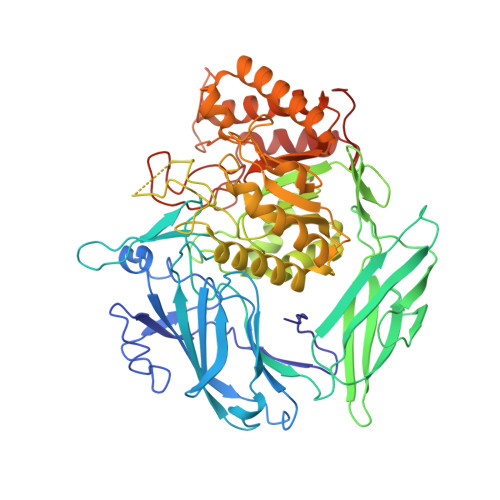
    3K46, 3K4A, 3K4D, 3LPF, 3LPG

  • PubMed Abstract: 

    The dose-limiting side effect of the common colon cancer chemotherapeutic CPT-11 is severe diarrhea caused by symbiotic bacterial β-glucuronidases that reactivate the drug in the gut. We sought to target these enzymes without killing the commensal bacteria essential for human health. Potent bacterial β-glucuronidase inhibitors were identified by high-throughput screening and shown to have no effect on the orthologous mammalian enzyme. Crystal structures established that selectivity was based on a loop unique to bacterial β-glucuronidases. Inhibitors were highly effective against the enzyme target in living aerobic and anaerobic bacteria, but did not kill the bacteria or harm mammalian cells. Finally, oral administration of an inhibitor protected mice from CPT-11-induced toxicity. Thus, drugs may be designed to inhibit undesirable enzyme activities in essential microbial symbiotes to enhance chemotherapeutic efficacy.


  • Organizational Affiliation
    • Department of Chemistry, University of North Carolina, Chapel Hill, NC 27599, USA.

Macromolecule Content 

  • Total Structure Weight: 137.49 kDa 
  • Atom Count: 9,918 
  • Modeled Residue Count: 1,193 
  • Deposited Residue Count: 1,210 
  • Unique protein chains: 1

Macromolecules

Find similar proteins by:|  3D Structure
Entity ID: 1
MoleculeChains  Sequence LengthOrganismDetailsImage
Beta-glucuronidase
A, B
605Escherichia coli K-12Mutation(s): 0 
Gene Names: b1617gurAgusAJW1609uidA
EC: 3.2.1.31
UniProt
Find proteins for P05804 (Escherichia coli (strain K12))
Explore P05804 
Go to UniProtKB:  P05804
Entity Groups
Sequence Clusters30% Identity50% Identity70% Identity90% Identity95% Identity100% Identity
UniProt GroupP05804
Sequence Annotations
Expand
Reference Sequence

Experimental Data & Validation

Experimental Data

  • Method: X-RAY DIFFRACTION
  • Resolution: 2.50 Å
  • R-Value Free:  0.267 (Depositor), 0.237 (DCC) 
  • R-Value Work:  0.214 (Depositor), 0.214 (DCC) 
  • R-Value Observed: 0.217 (Depositor) 
Space Group: C 1 2 1
Unit Cell:
Length ( Å )Angle ( ˚ )
a = 168.745α = 90
b = 76.193β = 125
c = 125.666γ = 90
Software Package:
Software NamePurpose
DENZOdata reduction
SCALEPACKdata scaling
PHASERphasing
PHENIXrefinement
PDB_EXTRACTdata extraction
HKL-2000data reduction
HKL-2000data scaling

Structure Validation

View Full Validation Report


Entry History 

Deposition Data

Revision History  (Full details and data files)

  • Version 1.0: 2010-11-17
    Type: Initial release
  • Version 1.1: 2011-07-13
    Changes: Source and taxonomy, Version format compliance
  • Version 1.2: 2017-11-01
    Changes: Refinement description
  • Version 1.3: 2023-09-06
    Changes: Data collection, Database references, Refinement description