3K3W | pdb_00003k3w

Thermostable Penicillin G acylase from Alcaligenes faecalis in orthorhombic form


Experimental Data Snapshot

  • Method: X-RAY DIFFRACTION
  • Resolution: 3.31 Å
  • R-Value Free: 
    0.315 (Depositor), 0.305 (DCC) 
  • R-Value Work: 
    0.281 (Depositor), 0.272 (DCC) 
  • R-Value Observed: 
    0.283 (Depositor) 

Starting Model: experimental
View more details

wwPDB Validation 3D Report Full Report

Validation slider image for 3K3W

This is version 1.5 of the entry. See complete history

Literature

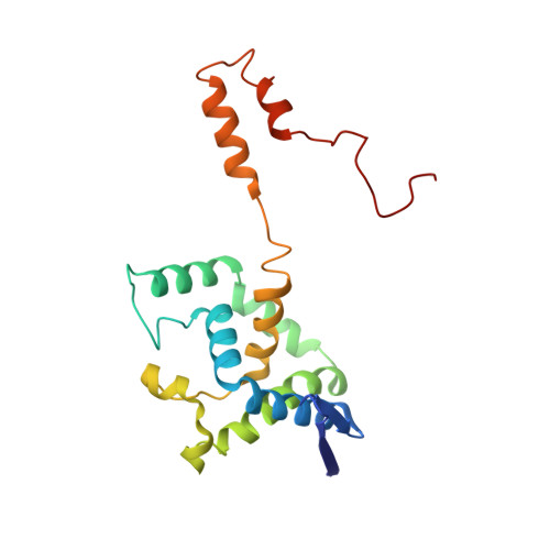
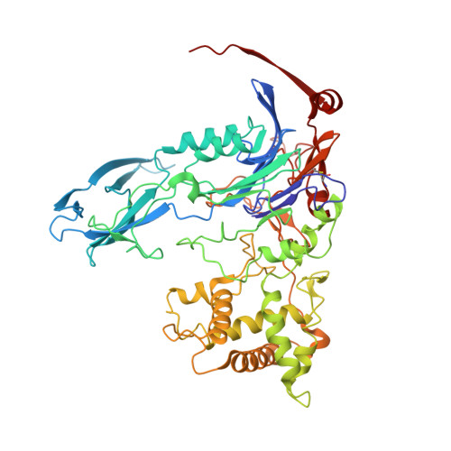
Crystallization and X-ray structure analysis of a thermostable penicillin G acylase from Alcaligenes faecalis.

Varshney, N.K.Kumar, R.S.Ignatova, Z.Prabhune, A.Pundle, A.Dodson, E.Suresh, C.G.

(2012) Acta Crystallogr Sect F Struct Biol Cryst Commun 68: 273-277

  • DOI: https://doi.org/10.1107/S1744309111053930
  • Primary Citation Related Structures: 
    3K3W, 3ML0

  • PubMed Abstract: 

    The enzyme penicillin G acylase (EC 3.5.1.11) catalyzes amide-bond cleavage in benzylpenicillin (penicillin G) to yield 6-aminopenicillanic acid, an intermediate chemical used in the production of semisynthetic penicillins. A thermostable penicillin G acylase from Alcaligenes faecalis (AfPGA) has been crystallized using the hanging-drop vapour-diffusion method in two different space groups: C222(1), with unit-cell parameters a = 72.9, b = 86.0, c = 260.2 , and P4(1)2(1)2, with unit-cell parameters a = b = 85.6, c = 298.8 . Data were collected at 293 and the structure was determined using the molecular-replacement method. Like other penicillin acylases, AfPGA belongs to the N-terminal nucleophilic hydrolase superfamily, has undergone post-translational processing and has a serine as the N-terminal residue of the β-chain. A disulfide bridge has been identified in the structure that was not found in the other two known penicillin G cylase structures. The presence of the disulfide bridge is perceived to be one factor that confers higher stability to this enzyme.


  • Organizational Affiliation
    • Division of Biochemical Sciences, National Chemical Laboratory, Pune 411 008, India.

Macromolecule Content 

  • Total Structure Weight: 85.16 kDa 
  • Atom Count: 6,011 
  • Modeled Residue Count: 747 
  • Deposited Residue Count: 747 
  • Unique protein chains: 2

Macromolecules

Find similar proteins by:|  3D Structure
Entity ID: 1
MoleculeChains  Sequence LengthOrganismDetailsImage
Penicillin G acylase196Alcaligenes faecalisMutation(s): 0 
Gene Names: pac
EC: 3.5.1.11
UniProt
Find proteins for O34142 (Alcaligenes faecalis)
Explore O34142 
Go to UniProtKB:  O34142
Entity Groups
Sequence Clusters30% Identity50% Identity70% Identity90% Identity95% Identity100% Identity
UniProt GroupO34142
Sequence Annotations
Expand
Reference Sequence
Find similar proteins by:|  3D Structure
Entity ID: 2
MoleculeChains  Sequence LengthOrganismDetailsImage
Penicillin G acylase551Alcaligenes faecalisMutation(s): 0 
Gene Names: pac
EC: 3.5.1.11
UniProt
Find proteins for O34142 (Alcaligenes faecalis)
Explore O34142 
Go to UniProtKB:  O34142
Entity Groups
Sequence Clusters30% Identity50% Identity70% Identity90% Identity95% Identity100% Identity
UniProt GroupO34142
Sequence Annotations
Expand
Reference Sequence

Small Molecules

Ligands 1 Unique
IDChains Name / Formula / InChI Key2D Diagram3D Interactions
CA

Query on CA


Download:Ideal Coordinates CCD File
C [auth B]CALCIUM ION
Ca
BHPQYMZQTOCNFJ-UHFFFAOYSA-N

Experimental Data & Validation

Experimental Data

  • Method: X-RAY DIFFRACTION
  • Resolution: 3.31 Å
  • R-Value Free:  0.315 (Depositor), 0.305 (DCC) 
  • R-Value Work:  0.281 (Depositor), 0.272 (DCC) 
  • R-Value Observed: 0.283 (Depositor) 
Space Group: C 2 2 21
Unit Cell:
Length ( Å )Angle ( ˚ )
a = 72.929α = 90
b = 86.016β = 90
c = 260.181γ = 90
Software Package:
Software NamePurpose
REFMACrefinement
PDB_EXTRACTdata extraction
CrystalCleardata collection
HKL-2000data reduction
HKL-2000data scaling
PHASERphasing

Structure Validation

View Full Validation Report


Entry History 

Deposition Data

Revision History  (Full details and data files)

  • Version 1.0: 2009-11-10
    Type: Initial release
  • Version 1.1: 2011-07-13
    Changes: Version format compliance
  • Version 1.2: 2012-03-14
    Changes: Database references, Structure summary
  • Version 1.3: 2020-01-01
    Changes: Advisory, Database references
  • Version 1.4: 2023-11-01
    Changes: Advisory, Data collection, Database references, Derived calculations, Refinement description
  • Version 1.5: 2024-11-20
    Changes: Structure summary