3K3T | pdb_00003k3t

E185A mutant of peptidoglycan hydrolase from Sphingomonas sp. A1


Experimental Data Snapshot

  • Method: X-RAY DIFFRACTION
  • Resolution: 1.75 Å
  • R-Value Free: 
    0.228 (Depositor), 0.227 (DCC) 
  • R-Value Work: 
    0.198 (Depositor), 0.198 (DCC) 
  • R-Value Observed: 
    0.199 (Depositor) 

Starting Model: experimental
View more details

wwPDB Validation 3D Report Full Report

Validation slider image for 3K3T

This is version 1.2 of the entry. See complete history

Literature

Mutational studies of the peptidoglycan hydrolase FlgJ of Sphingomonas sp. strain A1

Maruyama, Y.Ochiai, A.Itoh, T.Mikami, B.Hashimoto, W.Murata, K.

(2010) J Basic Microbiol 50: 311-317

  • DOI: https://doi.org/10.1002/jobm.200900249
  • Primary Citation Related Structures: 
    3K3T

  • PubMed Abstract: 

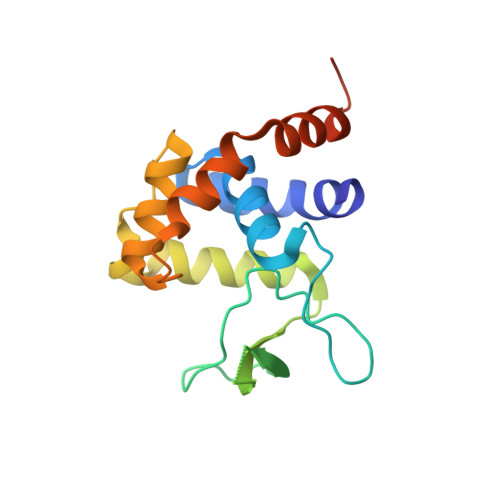
    The flagellar protein FlgJ, a member of glycoside hydrolase family 73, has N- and C-terminal domains that are responsible for flagellar rod assembly and peptidoglycan hydrolysis, respectively. The crystal structure of the C-terminal domain of SPH1045 (SPH1045-C), the FlgJ from Sphingomonas sp. strain A1, showed a long cleft formed by two lobes, alpha and beta. In this study, seven site-specific mutants of residues in the cleft were prepared and analyzed. Enzyme activity was reduced most significantly in mutants E185A and Y281A, followed by E224A. A comparison of the crystal structure of the inactive mutant E185A with that of other related enzymes revealed that Glu185 is structurally reasonable as the proton donor and that Tyr281 is close to Glu185. Glu224 is, however, far from the catalytic site, which is inconsistent with the decreased activity exhibited by E224A. The structural flexibility of Glu224 and its neighboring residues observed in SPH1045-C may indicate that this region is able to change its conformation upon substrate binding.


  • Organizational Affiliation
    • Laboratory of Basic and Applied Molecular Biotechnology, Graduate School of Agriculture, Kyoto University, Uji, Kyoto, Japan.

Macromolecule Content 

  • Total Structure Weight: 18.86 kDa 
  • Atom Count: 1,317 
  • Modeled Residue Count: 152 
  • Deposited Residue Count: 170 
  • Unique protein chains: 1

Macromolecules

Find similar proteins by:|  3D Structure
Entity ID: 1
MoleculeChains  Sequence LengthOrganismDetailsImage
Peptidoglycan hydrolase FlgJ170Sphingomonas sp. A1Mutation(s): 3 
Gene Names: flgJ
EC: 3.2.1
UniProt
Find proteins for B7XH69 (Sphingomonas sp. A1)
Explore B7XH69 
Go to UniProtKB:  B7XH69
Entity Groups
Sequence Clusters30% Identity50% Identity70% Identity90% Identity95% Identity100% Identity
UniProt GroupB7XH69
Sequence Annotations
Expand
Reference Sequence

Experimental Data & Validation

Experimental Data

  • Method: X-RAY DIFFRACTION
  • Resolution: 1.75 Å
  • R-Value Free:  0.228 (Depositor), 0.227 (DCC) 
  • R-Value Work:  0.198 (Depositor), 0.198 (DCC) 
  • R-Value Observed: 0.199 (Depositor) 
Space Group: P 43 21 2
Unit Cell:
Length ( Å )Angle ( ˚ )
a = 54.441α = 90
b = 54.441β = 90
c = 101.569γ = 90
Software Package:
Software NamePurpose
MOLREPphasing
REFMACrefinement
HKL-2000data reduction
HKL-2000data scaling

Structure Validation

View Full Validation Report


Entry History 

Deposition Data

Revision History  (Full details and data files)

  • Version 1.0: 2010-07-14
    Type: Initial release
  • Version 1.1: 2011-07-13
    Changes: Version format compliance
  • Version 1.2: 2023-11-01
    Changes: Data collection, Database references, Derived calculations, Refinement description