


Crystal Structure of Spin Labeled T4 Lysozyme Mutant K65V1/R76V1
Toledo Warshaviak, D., Cascio, D., Khramtsov, V.V., Hubbell, W.L.To be published.
Experimental Data Snapshot
Starting Model: experimental
View more details
Entity ID: 1 | |||||
|---|---|---|---|---|---|
| Molecule | Chains | Sequence Length | Organism | Details | Image |
| Lysozyme | 164 | Tequatrovirus T4 | Mutation(s): 4 Gene Names: E, Lysozyme EC: 3.2.1.17 | 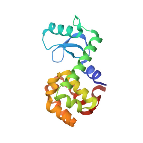 | |
UniProt | |||||
Entity Groups | |||||
| Sequence Clusters | 30% Identity50% Identity70% Identity90% Identity95% Identity100% Identity | ||||
| UniProt Group | P00720 | ||||
Sequence AnnotationsExpand | |||||
Reference Sequence | |||||
| Ligands 4 Unique | |||||
|---|---|---|---|---|---|
| ID | Chains | Name / Formula / InChI Key | 2D Diagram | 3D Interactions | |
| V1A Download:Ideal Coordinates CCD File | B [auth A], D [auth A] | S-(1-oxyl-2,2,5,5-tetramethyl-2,5-dihydro-1H-imidazol-4-yl) methanesulfonothioate C8 H15 N2 O3 S2 GGUBULNJTWFONN-UHFFFAOYSA-N |  | ||
| HEZ Download:Ideal Coordinates CCD File | C [auth A], E [auth A] | HEXANE-1,6-DIOL C6 H14 O2 XXMIOPMDWAUFGU-UHFFFAOYSA-N |  | ||
| K Download:Ideal Coordinates CCD File | H [auth A] | POTASSIUM ION K NPYPAHLBTDXSSS-UHFFFAOYSA-N |  | ||
| CL Download:Ideal Coordinates CCD File | F [auth A], G [auth A] | CHLORIDE ION Cl VEXZGXHMUGYJMC-UHFFFAOYSA-M |  | ||
| Length ( Å ) | Angle ( ˚ ) |
|---|---|
| a = 59.547 | α = 90 |
| b = 59.547 | β = 90 |
| c = 95.28 | γ = 120 |
| Software Name | Purpose |
|---|---|
| DENZO | data reduction |
| SCALEPACK | data scaling |
| PHASER | phasing |
| PHENIX | refinement |
| PDB_EXTRACT | data extraction |


