3K2B | pdb_00003k2b

Crystal structure of photosynthetic A4 isoform glyceraldehyde-3-phosphate dehydrogenase complexed with NAD, from Arabidopsis thaliana


Experimental Data Snapshot

  • Method: X-RAY DIFFRACTION
  • Resolution: 2.60 Å
  • R-Value Free: 
    0.289 (Depositor), 0.288 (DCC) 
  • R-Value Work: 
    0.237 (Depositor), 0.237 (DCC) 
  • R-Value Observed: 
    0.237 (Depositor) 

Starting Model: experimental
View more details

wwPDB Validation 3D Report Full Report

Validation slider image for 3K2B

Ligand Structure Quality Assessment 


This is version 1.3 of the entry. See complete history

Literature

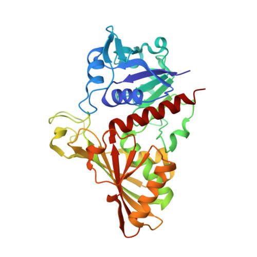
Structure of photosynthetic glyceraldehyde-3-phosphate dehydrogenase (isoform A4) from Arabidopsis thaliana in complex with NAD

Fermani, S.Sparla, F.Marri, L.Thumiger, A.Pupillo, P.Falini, G.Trost, P.

(2010) Acta Crystallogr Sect F Struct Biol Cryst Commun 66: 621-626

  • DOI: https://doi.org/10.1107/S1744309110013527
  • Primary Citation Related Structures: 
    3K2B

  • PubMed Abstract: 

    The crystal structure of the A(4) isoform of photosynthetic glyceraldehyde-3-phosphate dehydrogenase (GAPDH) from Arabidopsis thaliana, expressed in recombinant form and complexed with NAD, is reported. The crystals, which were grown in 2.4 M ammonium sulfate and 0.1 M sodium citrate, belonged to space group I222. The asymmetric unit includes ten subunits, i.e. two independent tetramers plus a dimer that generates a third tetramer by a crystallographic symmetry operation. The crystal structure was solved by molecular replacement and refined to an R factor of 23.7% and an R(free) factor of 28.9% at 2.6 A resolution. In the final model, each subunit binds one NAD(+) molecule and two sulfates, which occupy the P(s) and the P(i) anion-binding sites. Detailed knowledge of this structure is instrumental for structural investigation of supramolecular complexes of A(4)-GAPDH, phosphoribulokinase and CP12, which are involved in the regulation of photosynthesis in the model plant A. thaliana.


  • Organizational Affiliation
    • Department of Chemistry, University of Bologna, Via Selmi 2, 40126 Bologna, Italy.

Macromolecule Content 

  • Total Structure Weight: 374.87 kDa 
  • Atom Count: 27,072 
  • Modeled Residue Count: 3,363 
  • Deposited Residue Count: 3,370 
  • Unique protein chains: 1

Macromolecules

Find similar proteins by:|  3D Structure
Entity ID: 1
MoleculeChains  Sequence LengthOrganismDetailsImage
Glyceraldehyde-3-phosphate dehydrogenase A, chloroplastic337Arabidopsis thalianaMutation(s): 0 
Gene Names: GAPA
EC: 1.2.1.13
UniProt
Find proteins for P25856 (Arabidopsis thaliana)
Explore P25856 
Go to UniProtKB:  P25856
Entity Groups
Sequence Clusters30% Identity50% Identity70% Identity90% Identity95% Identity100% Identity
UniProt GroupP25856
Sequence Annotations
Expand
Reference Sequence

Small Molecules

Ligands 2 Unique
IDChains Name / Formula / InChI Key2D Diagram3D Interactions
NAD

Query on NAD


Download:Ideal Coordinates CCD File
AB [auth O]
DA [auth D]
HA [auth E]
HB [auth Q]
K [auth A]
AB [auth O],
DA [auth D],
HA [auth E],
HB [auth Q],
K [auth A],
MA [auth F],
QA [auth G],
S [auth B],
WA [auth H],
Y [auth C]
NICOTINAMIDE-ADENINE-DINUCLEOTIDE
C21 H27 N7 O14 P2
BAWFJGJZGIEFAR-NNYOXOHSSA-N
SO4

Query on SO4


Download:Ideal Coordinates CCD File
AA [auth C]
BA [auth C]
BB [auth O]
CA [auth C]
CB [auth O]
AA [auth C],
BA [auth C],
BB [auth O],
CA [auth C],
CB [auth O],
DB [auth O],
EA [auth D],
EB [auth O],
FA [auth D],
FB [auth O],
GA [auth D],
GB [auth O],
IA [auth E],
IB [auth Q],
JA [auth E],
JB [auth Q],
KA [auth E],
KB [auth Q],
L [auth A],
LA [auth E],
LB [auth Q],
M [auth A],
MB [auth Q],
N [auth A],
NA [auth F],
O [auth A],
OA [auth F],
P [auth A],
PA [auth F],
Q [auth A],
R [auth A],
RA [auth G],
SA [auth G],
T [auth B],
TA [auth G],
U [auth B],
UA [auth G],
V [auth B],
VA [auth G],
W [auth B],
X [auth B],
XA [auth H],
YA [auth H],
Z [auth C],
ZA [auth H]
SULFATE ION
O4 S
QAOWNCQODCNURD-UHFFFAOYSA-L

Experimental Data & Validation

Experimental Data

  • Method: X-RAY DIFFRACTION
  • Resolution: 2.60 Å
  • R-Value Free:  0.289 (Depositor), 0.288 (DCC) 
  • R-Value Work:  0.237 (Depositor), 0.237 (DCC) 
  • R-Value Observed: 0.237 (Depositor) 
Space Group: I 2 2 2
Unit Cell:
Length ( Å )Angle ( ˚ )
a = 150.738α = 90
b = 188.625β = 90
c = 314.129γ = 90
Software Package:
Software NamePurpose
DNAdata collection
EPMRphasing
CNSrefinement
DENZOdata reduction
SCALEPACKdata scaling

Structure Validation

View Full Validation Report


Ligand Structure Quality Assessment 


Entry History 

Deposition Data

Revision History  (Full details and data files)

  • Version 1.0: 2010-06-09
    Type: Initial release
  • Version 1.1: 2011-07-13
    Changes: Version format compliance
  • Version 1.2: 2012-05-02
    Changes: Database references
  • Version 1.3: 2023-11-01
    Changes: Data collection, Database references, Derived calculations, Refinement description