3JZI | pdb_00003jzi

Crystal structure of biotin carboxylase from E. Coli in complex with benzimidazole series


Experimental Data Snapshot

  • Method: X-RAY DIFFRACTION
  • Resolution: 2.31 Å
  • R-Value Free: 
    0.223 (Depositor), 0.220 (DCC) 
  • R-Value Work: 
    0.187 (Depositor), 0.183 (DCC) 

Starting Model: experimental
View more details

wwPDB Validation 3D Report Full Report

Validation slider image for 3JZI

Ligand Structure Quality Assessment 


This is version 1.2 of the entry. See complete history

Literature

Discovery and optimization of antibacterial AccC inhibitors.

Cheng, C.C.Shipps, G.W.Yang, Z.Sun, B.Kawahata, N.Soucy, K.A.Soriano, A.Orth, P.Xiao, L.Mann, P.Black, T.

(2009) Bioorg Med Chem Lett 19: 6507-6514

  • DOI: https://doi.org/10.1016/j.bmcl.2009.10.057
  • Primary Citation Related Structures: 
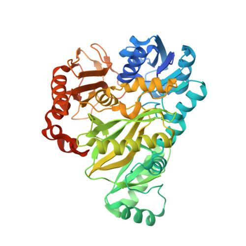
    3JZF, 3JZI

  • PubMed Abstract: 

    The biotin carboxylase (AccC) is part of the multi-component bacterial acetyl coenzyme-A carboxylase (ACCase) and is essential for pathogen survival. We describe herein the affinity optimization of an initial hit to give 2-(2-chlorobenzylamino)-1-(cyclohexylmethyl)-1H-benzo[d]imidazole-5-carboxamide (1), which was identified using our proprietary Automated Ligand Identification System (ALIS).(1) The X-ray co-crystal structure of 1 was solved and revealed several key interactions and opportunities for further optimization in the ATP site of AccC. Structure Based Drug Design (SBDD) and parallel synthetic approaches resulted in a novel series of AccC inhibitors, exemplified by (R)-2-(2-chlorobenzylamino)-1-(2,3-dihydro-1H-inden-1-yl)-1H-imidazo[4,5-b]pyridine-5-carboxamide (40). This compound is a potent and selective inhibitor of bacterial AccC with an IC(50) of 20 nM and a MIC of 0.8 microg/mL against a sensitized strain of Escherichia coli (HS294 E. coli).


  • Organizational Affiliation
    • Schering-Plough Research Institute, Cambridge, MA 02141, United States. cccheng@alum.mit.edu

Macromolecule Content 

  • Total Structure Weight: 107.9 kDa 
  • Atom Count: 7,280 
  • Modeled Residue Count: 891 
  • Deposited Residue Count: 972 
  • Unique protein chains: 1

Macromolecules

Find similar proteins by:|  3D Structure
Entity ID: 1
MoleculeChains  Sequence LengthOrganismDetailsImage
Biotin carboxylase
A, B
486Escherichia coli K-12Mutation(s): 0 
Gene Names: accCfabGb3256JW3224
EC: 6.3.4.14 (PDB Primary Data), 6.4.1.2 (PDB Primary Data)
UniProt
Find proteins for P24182 (Escherichia coli (strain K12))
Explore P24182 
Go to UniProtKB:  P24182
Entity Groups
Sequence Clusters30% Identity50% Identity70% Identity90% Identity95% Identity100% Identity
UniProt GroupP24182
Sequence Annotations
Expand
Reference Sequence

Small Molecules

Ligands 1 Unique
IDChains Name / Formula / InChI Key2D Diagram3D Interactions
JZL

Query on JZL


Download:Ideal Coordinates CCD File
C [auth A],
D [auth B]
7-amino-2-[(2-chlorobenzyl)amino]-1-{[(1S,2S)-2-hydroxycycloheptyl]methyl}-1H-benzimidazole-5-carboxamide
C23 H28 Cl N5 O2
AQAQDMCJHAROPH-YWZLYKJASA-N

Experimental Data & Validation

Experimental Data

  • Method: X-RAY DIFFRACTION
  • Resolution: 2.31 Å
  • R-Value Free:  0.223 (Depositor), 0.220 (DCC) 
  • R-Value Work:  0.187 (Depositor), 0.183 (DCC) 
Space Group: P 21 21 21
Unit Cell:
Length ( Å )Angle ( ˚ )
a = 84.758α = 90
b = 107.816β = 90
c = 121.941γ = 90
Software Package:
Software NamePurpose
CrystalCleardata collection
AMoREphasing
BUSTERrefinement
DENZOdata reduction
HKL-2000data scaling

Structure Validation

View Full Validation Report


Ligand Structure Quality Assessment 


Entry History 

Deposition Data

Revision History  (Full details and data files)

  • Version 1.0: 2009-11-03
    Type: Initial release
  • Version 1.1: 2011-07-13
    Changes: Version format compliance
  • Version 1.2: 2023-09-06
    Changes: Data collection, Database references, Derived calculations, Refinement description