3JW1 | pdb_00003jw1

Crystal Structure of Bovine Pancreatic Ribonuclease Complexed with Uridine-5'-monophosphate at 1.60 A Resolution


Experimental Data Snapshot

  • Method: X-RAY DIFFRACTION
  • Resolution: 1.60 Å
  • R-Value Free: 
    0.253 (Depositor), 0.277 (DCC) 
  • R-Value Work: 
    0.197 (Depositor), 0.219 (DCC) 
  • R-Value Observed: 
    0.203 (Depositor) 

Starting Model: experimental
View more details

wwPDB Validation 3D Report Full Report

Validation slider image for 3JW1

Ligand Structure Quality Assessment 


This is version 1.4 of the entry. See complete history

Literature

Structure of bovine pancreatic ribonuclease complexed with uridine 5'-monophosphate at 1.60 A resolution.

Larson, S.B.Day, J.S.Nguyen, C.Cudney, R.McPherson, A.

(2010) Acta Crystallogr Sect F Struct Biol Cryst Commun 66: 113-120

  • DOI: https://doi.org/10.1107/S174430910905194X
  • Primary Citation Related Structures: 
    3JW1

  • PubMed Abstract: 

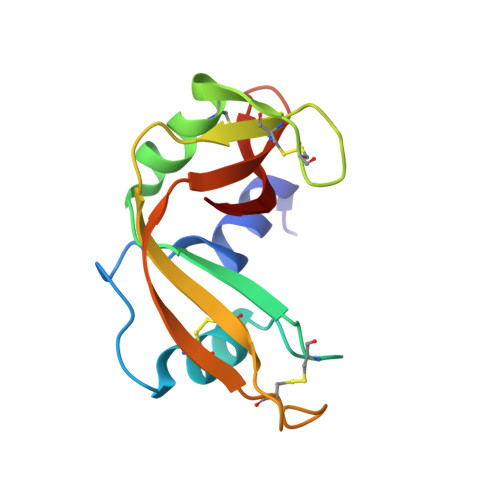
    Bovine pancreatic ribonuclease A (RNase A) was crystallized from a mixture of small molecules containing basic fuchsin, tobramycin and uridine 5'-monophosphate (U5P). Solution of the crystal structure revealed that the enzyme was selectively bound to U5P, with the pyrimidine ring of U5P residing in the pyrimidine-binding site at Thr45. The structure was refined to an R factor of 0.197 and an R(free) of 0.253.


  • Organizational Affiliation
    • Department of Molecular Biology and Biochemistry, The University of California, Irvine, CA 92697-3900, USA.

Macromolecule Content 

  • Total Structure Weight: 28.07 kDa 
  • Atom Count: 2,203 
  • Modeled Residue Count: 248 
  • Deposited Residue Count: 248 
  • Unique protein chains: 1

Macromolecules

Find similar proteins by:|  3D Structure
Entity ID: 1
MoleculeChains  Sequence LengthOrganismDetailsImage
Ribonuclease pancreatic
A, B
124Bos taurusMutation(s): 0 
EC: 3.1.27.5 (PDB Primary Data), 4.6.1.18 (UniProt)
UniProt
Find proteins for P61823 (Bos taurus)
Explore P61823 
Go to UniProtKB:  P61823
Entity Groups
Sequence Clusters30% Identity50% Identity70% Identity90% Identity95% Identity100% Identity
UniProt GroupP61823
Sequence Annotations
Expand
Reference Sequence

Experimental Data & Validation

Experimental Data

  • Method: X-RAY DIFFRACTION
  • Resolution: 1.60 Å
  • R-Value Free:  0.253 (Depositor), 0.277 (DCC) 
  • R-Value Work:  0.197 (Depositor), 0.219 (DCC) 
  • R-Value Observed: 0.203 (Depositor) 
Space Group: P 1 21 1
Unit Cell:
Length ( Å )Angle ( ˚ )
a = 30.73α = 90
b = 74.85β = 107.8
c = 50.52γ = 90
Software Package:
Software NamePurpose
REFMACrefinement
d*TREKdata reduction
d*TREKdata scaling

Structure Validation

View Full Validation Report


Ligand Structure Quality Assessment 


Entry History 

Deposition Data

Revision History  (Full details and data files)

  • Version 1.0: 2009-10-06
    Type: Initial release
  • Version 1.1: 2011-07-13
    Changes: Advisory, Refinement description, Version format compliance
  • Version 1.2: 2017-11-01
    Changes: Refinement description
  • Version 1.3: 2023-09-06
    Changes: Data collection, Database references, Derived calculations, Refinement description
  • Version 1.4: 2024-11-20
    Changes: Structure summary