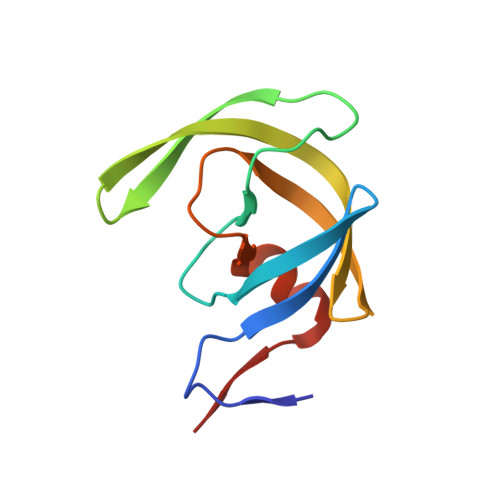
3JVY

HIV-1 Protease Mutant G86A with DARUNAVIR


Experimental Data Snapshot

  • Method: X-RAY DIFFRACTION
  • Resolution: 1.60 Å
  • R-Value Free: 0.256 
  • R-Value Work: 0.186 
  • R-Value Observed: 0.186 

Starting Model: experimental
View more details

wwPDB Validation   3D Report Full Report


Ligand Structure Quality Assessment 


This is version 1.4 of the entry. See complete history


Literature

Highly conserved glycine 86 and arginine 87 residues contribute differently to the structure and activity of the mature HIV-1 protease

Ishima, R.Gong, Q.Tie, Y.Weber, I.T.Louis, J.M.

(2009) Proteins 78: 1015-1025

  • DOI: https://doi.org/10.1002/prot.22625
  • Primary Citation of Related Structures:  
    3JVW, 3JVY, 3JW2

  • PubMed Abstract: 

    The structural and functional role of conserved residue G86 in HIV-1 protease (PR) was investigated by NMR and crystallographic analyses of substitution mutations of glycine to alanine and serine (PR(G86A) and PR(G86S)). While PR(G86S) had undetectable catalytic activity, PR(G86A) exhibited approximately 6000-fold lower catalytic activity than PR. (1)H-(15)N NMR correlation spectra revealed that PR(G86A) and PR(G86S) are dimeric, exhibiting dimer dissociation constants (K(d)) of approximately 0.5 and approximately 3.2 muM, respectively, which are significantly lower than that seen for PR with R87K mutation (K(d) > 1 mM). Thus, the G86 mutants, despite being partially dimeric under the assay conditions, are defective in catalyzing substrate hydrolysis. NMR spectra revealed no changes in the chemical shifts even in the presence of excess substrate, indicating very poor binding of the substrate. Both NMR chemical shift data and crystal structures of PR(G86A) and PR(G86S) in the presence of active-site inhibitors indicated high structural similarity to previously described PR/inhibitor complexes, except for specific perturbations within the active site loop and around the mutation site. The crystal structures in the presence of the inhibitor showed that the region around residue 86 was connected to the active site by a conserved network of hydrogen bonds, and the two regions moved further apart in the mutants. Overall, in contrast to the role of R87 in contributing significantly to the dimer stability of PR, G86 is likely to play an important role in maintaining the correct geometry of the active site loop in the PR dimer for substrate binding and hydrolysis. Proteins 2010. (c) 2009 Wiley-Liss, Inc.


  • Organizational Affiliation

    Department of Structural Biology, University of Pittsburgh School of Medicine, Pittsburgh, Pennsylvania 15260, USA. ishima@pitt.edu


Macromolecules
Find similar proteins by:  (by identity cutoff)  |  3D Structure
Entity ID: 1
MoleculeChains Sequence LengthOrganismDetailsImage
Gag-Pol polyprotein
A, B
99Human immunodeficiency virus type 1 (BRU ISOLATE)Mutation(s): 6 
Gene Names: gag-polHuman immunodeficiency virus type 1
EC: 3.4.23.16
UniProt
Find proteins for P03367 (Human immunodeficiency virus type 1 group M subtype B (isolate BRU/LAI))
Explore P03367 
Go to UniProtKB:  P03367
Entity Groups  
Sequence Clusters30% Identity50% Identity70% Identity90% Identity95% Identity100% Identity
UniProt GroupP03367
Sequence Annotations
Expand
  • Reference Sequence
Small Molecules
Ligands 3 Unique
IDChains Name / Formula / InChI Key2D Diagram3D Interactions
017
Query on 017

Download Ideal Coordinates CCD File 
E [auth B](3R,3AS,6AR)-HEXAHYDROFURO[2,3-B]FURAN-3-YL(1S,2R)-3-[[(4-AMINOPHENYL)SULFONYL](ISOBUTYL)AMINO]-1-BENZYL-2-HYDROXYPROPYLCARBAMATE
C27 H37 N3 O7 S
CJBJHOAVZSMMDJ-HEXNFIEUSA-N
CL
Query on CL

Download Ideal Coordinates CCD File 
D [auth A],
F [auth B]
CHLORIDE ION
Cl
VEXZGXHMUGYJMC-UHFFFAOYSA-M
NA
Query on NA

Download Ideal Coordinates CCD File 
C [auth A]SODIUM ION
Na
FKNQFGJONOIPTF-UHFFFAOYSA-N
Experimental Data & Validation

Experimental Data

  • Method: X-RAY DIFFRACTION
  • Resolution: 1.60 Å
  • R-Value Free: 0.256 
  • R-Value Work: 0.186 
  • R-Value Observed: 0.186 
  • Space Group: P 21 21 2
Unit Cell:
Length ( Å )Angle ( ˚ )
a = 58.328α = 90
b = 86.433β = 90
c = 46.204γ = 90
Software Package:
Software NamePurpose
MAR345data collection
AMoREphasing
SHELXL-97refinement
HKL-2000data reduction
HKL-2000data scaling

Structure Validation

View Full Validation Report


Ligand Structure Quality Assessment 


Entry History 

Deposition Data

Revision History  (Full details and data files)

  • Version 1.0: 2009-12-08
    Type: Initial release
  • Version 1.1: 2011-07-13
    Changes: Non-polymer description, Version format compliance
  • Version 1.2: 2017-11-01
    Changes: Refinement description
  • Version 1.3: 2021-10-13
    Changes: Database references, Derived calculations, Structure summary
  • Version 1.4: 2023-09-06
    Changes: Data collection, Refinement description