3JV3 | pdb_00003jv3

Structure of SH3E-DH unit of murine intersectin-1L


Experimental Data Snapshot

  • Method: X-RAY DIFFRACTION
  • Resolution: 2.40 Å
  • R-Value Free: 
    0.287 (Depositor), 0.286 (DCC) 
  • R-Value Work: 
    0.248 (Depositor), 0.247 (DCC) 
  • R-Value Observed: 
    0.250 (Depositor) 

wwPDB Validation 3D Report Full Report

Validation slider image for 3JV3

This is version 1.3 of the entry. See complete history

Literature

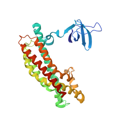
The minimal autoinhibited unit of the guanine nucleotide exchange factor intersectin.

Ahmad, K.F.Lim, W.A.

(2010) PLoS One 5: e11291-e11291

  • DOI: https://doi.org/10.1371/journal.pone.0011291
  • Primary Citation Related Structures: 
    3JV3

  • PubMed Abstract: 

    Intersectin-1L is a member of the Dbl homology (DH) domain guanine nucleotide exchange factors (GEF) which control Rho-family GTPase signaling. Intersectin-1L is a GEF that is specific for Cdc42. It plays an important role in endocytosis, and is regulated by several partners including the actin regulator N-WASP. Intact intersectin-1L shows low Cdc42 exchange activity, although the isolated catalytic DH domain shows high activity. This finding suggests that the molecule is autoinhibited. To investigate the mechanism of autoinhibition we have constructed a series of domain deletions. We find that the five SH3 domains of intersectin are important for autoinhibition, with the fifth domain (SH3(E)) being sufficient for the bulk of the autoinhibitory effect. This SH3 domain appears to primarily interact with the DH domain. We have determined the crystal structure of the SH3(E)-DH domain construct, which shows a domain swapped arrangement in which the SH3 from one monomer interacts with the DH domain of the other monomer. Analytical ultracentrifugation and gel filtration, however, show that under biochemical concentrations, the construct is fully monomeric. Thus we propose that the actual autoinhibited structure contains the related intramolecular SH3(E)-DH interaction. We propose a model in which this intramolecular interaction may block or distort the GTPase binding region of the DH domain.


  • Organizational Affiliation
    • Department of Cellular and Molecular Pharmacology, University of California San Francisco, San Francisco, California, United States of America.

Macromolecule Content 

  • Total Structure Weight: 65.44 kDa 
  • Atom Count: 4,129 
  • Modeled Residue Count: 502 
  • Deposited Residue Count: 566 
  • Unique protein chains: 1

Macromolecules

Find similar proteins by:|  3D Structure
Entity ID: 1
MoleculeChains  Sequence LengthOrganismDetailsImage
Intersectin-1
A, B
283Mus musculusMutation(s): 0 
Gene Names: Itsn1Ese1Itsn
UniProt & NIH Common Fund Data Resources
Find proteins for Q9Z0R4 (Mus musculus)
Explore Q9Z0R4 
Go to UniProtKB:  Q9Z0R4
IMPC:  MGI:1338069
Entity Groups
Sequence Clusters30% Identity50% Identity70% Identity90% Identity95% Identity100% Identity
UniProt GroupQ9Z0R4
Sequence Annotations
Expand
Reference Sequence

Experimental Data & Validation

Experimental Data

  • Method: X-RAY DIFFRACTION
  • Resolution: 2.40 Å
  • R-Value Free:  0.287 (Depositor), 0.286 (DCC) 
  • R-Value Work:  0.248 (Depositor), 0.247 (DCC) 
  • R-Value Observed: 0.250 (Depositor) 
Space Group: P 32 2 1
Unit Cell:
Length ( Å )Angle ( ˚ )
a = 67.044α = 90
b = 67.044β = 90
c = 341.818γ = 120
Software Package:
Software NamePurpose
DENZOdata reduction
SCALEPACKdata scaling
PHASERphasing
CNSrefinement
PDB_EXTRACTdata extraction
ADSCdata collection

Structure Validation

View Full Validation Report


Entry History 

Deposition Data

  • Released Date: 2010-07-14 
  • Deposition Author(s): Ahmad, K.F.

Revision History  (Full details and data files)

  • Version 1.0: 2010-07-14
    Type: Initial release
  • Version 1.1: 2011-07-13
    Changes: Version format compliance
  • Version 1.2: 2017-11-01
    Changes: Refinement description
  • Version 1.3: 2024-02-21
    Changes: Data collection, Database references