3JRN | pdb_00003jrn

Crystal structure of TIR domain from Arabidopsis Thaliana


Experimental Data Snapshot

  • Method: X-RAY DIFFRACTION
  • Resolution: 2.00 Å
  • R-Value Free: 
    0.208 (Depositor), 0.214 (DCC) 
  • R-Value Work: 
    0.168 (Depositor), 0.182 (DCC) 

wwPDB Validation 3D Report Full Report

Validation slider image for 3JRN

This is version 1.2 of the entry. See complete history

Literature

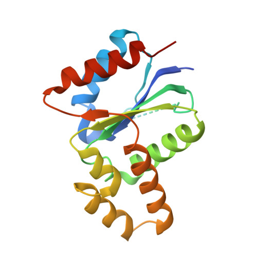
The crystal structure of a TIR domain from Arabidopsis thaliana reveals a conserved helical region unique to plants.

Chan, S.L.Mukasa, T.Santelli, E.Low, L.Y.Pascual, J.

(2009) Protein Sci 19: 155-161

  • DOI: https://doi.org/10.1002/pro.275
  • Primary Citation Related Structures: 
    3JRN

  • PubMed Abstract: 

    Plants use a highly evolved immune system to exhibit defense response against microbial infections. The plant TIR domain, together with the nucleotide-binding (NB) domain and/or a LRR region, forms a type of molecule, named resistance (R) proteins, that interact with microbial effector proteins and elicit hypersensitive responses against infection. Here, we report the first crystal structure of a plant TIR domain from Arabidopsis thaliana (AtTIR) solved at a resolution of 2.0 A. The structure consists of five beta-strands forming a parallel beta-sheet at the core of the protein. The beta-strands are connected by a series of alpha-helices and the overall fold mimics closely that of other mammalian and bacterial TIR domains. However, the region of the alphaD-helix reveals significant differences when compared with other TIR structures, especially the alphaD3-helix that corresponds to an insertion only present in plant TIR domains. Available mutagenesis data suggest that several conserved and exposed residues in this region are involved in the plant TIR signaling function.


  • Organizational Affiliation
    • Infectious Diseases Center, Burnham Institute for Medical Research, La Jolla, California 92037, USA.

Macromolecule Content 

  • Total Structure Weight: 20.35 kDa 
  • Atom Count: 1,357 
  • Modeled Residue Count: 151 
  • Deposited Residue Count: 176 
  • Unique protein chains: 1

Macromolecules

Find similar proteins by:|  3D Structure
Entity ID: 1
MoleculeChains  Sequence LengthOrganismDetailsImage
AT1G72930 protein176Arabidopsis thalianaMutation(s): 0 
Gene Names: AT1G72930At1g72930/F3N23.13AtTIRF3N23.13
UniProt
Find proteins for Q9SSN3 (Arabidopsis thaliana)
Explore Q9SSN3 
Go to UniProtKB:  Q9SSN3
Entity Groups
Sequence Clusters30% Identity50% Identity70% Identity90% Identity95% Identity100% Identity
UniProt GroupQ9SSN3
Sequence Annotations
Expand
Reference Sequence

Small Molecules

Ligands 1 Unique
IDChains Name / Formula / InChI Key2D Diagram3D Interactions
ARS

Query on ARS


Download:Ideal Coordinates CCD File
B [auth A]ARSENIC
As
RBFQJDQYXXHULB-UHFFFAOYSA-N

Experimental Data & Validation

Experimental Data

  • Method: X-RAY DIFFRACTION
  • Resolution: 2.00 Å
  • R-Value Free:  0.208 (Depositor), 0.214 (DCC) 
  • R-Value Work:  0.168 (Depositor), 0.182 (DCC) 
Space Group: P 41 21 2
Unit Cell:
Length ( Å )Angle ( ˚ )
a = 71.21α = 90
b = 71.21β = 90
c = 60.27γ = 90
Software Package:
Software NamePurpose
CNSrefinement
PDB_EXTRACTdata extraction
HKL-2000data collection
HKL-2000data reduction
HKL-2000data scaling
SOLVEphasing

Structure Validation

View Full Validation Report


Entry History 

Deposition Data

Revision History  (Full details and data files)

  • Version 1.0: 2009-10-20
    Type: Initial release
  • Version 1.1: 2011-07-13
    Changes: Version format compliance
  • Version 1.2: 2024-02-21
    Changes: Data collection, Database references, Derived calculations