3JBR | pdb_00003jbr

Cryo-EM structure of the rabbit voltage-gated calcium channel Cav1.1 complex at 4.2 angstrom


Experimental Data Snapshot

  • Method: ELECTRON MICROSCOPY
  • Resolution: 4.20 Å
  • Aggregation State: PARTICLE 
  • Reconstruction Method: SINGLE PARTICLE 

Starting Model: experimental
View more details

wwPDB Validation 3D Report Full Report

Validation slider image for 3JBR

This is version 1.3 of the entry. See complete history

Literature

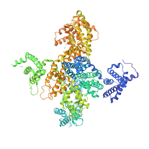
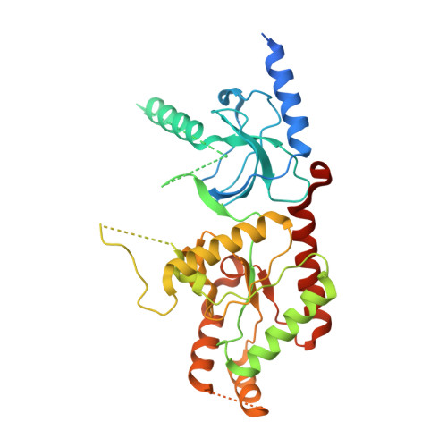
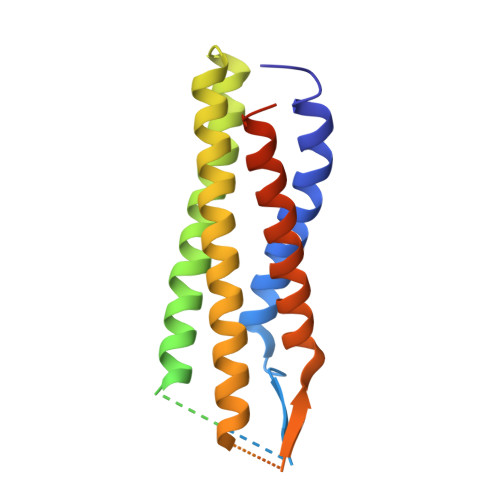
Structure of the voltage-gated calcium channel Cav1.1 complex

Wu, J.P.Yan, Z.Li, Z.Q.Yan, C.G.Lu, S.Dong, M.Q.Yan, N.

(2015) Science 350: aad2395-aad2395

  • DOI: https://doi.org/10.1126/science.aad2395
  • Primary Citation Related Structures: 
    3JBR

  • PubMed Abstract: 

    The voltage-gated calcium channel Ca(v)1.1 is engaged in the excitation-contraction coupling of skeletal muscles. The Ca(v)1.1 complex consists of the pore-forming subunit α1 and auxiliary subunits α2δ, β, and γ. We report the structure of the rabbit Ca(v)1.1 complex determined by single-particle cryo-electron microscopy. The four homologous repeats of the α1 subunit are arranged clockwise in the extracellular view. The γ subunit, whose structure resembles claudins, interacts with the voltage-sensing domain of repeat IV (VSD(IV)), whereas the cytosolic β subunit is located adjacent to VSD(II) of α1. The α2 subunit interacts with the extracellular loops of repeats I to III through its VWA and Cache1 domains. The structure reveals the architecture of a prototypical eukaryotic Ca(v) channel and provides a framework for understanding the function and disease mechanisms of Ca(v) and Na(v) channels.


  • Organizational Affiliation
    • State Key Laboratory of Membrane Biology, Tsinghua University, Beijing 100084, China. Tsinghua-Peking Joint Center for Life Sciences, Tsinghua University, Beijing 100084, China. Center for Structural Biology, School of Life Sciences and School of Medicine, Tsinghua University, Beijing 100084, China.

Macromolecule Content 

  • Total Structure Weight: 394.53 kDa 
  • Atom Count: 16,467 
  • Modeled Residue Count: 2,515 
  • Deposited Residue Count: 3,557 
  • Unique protein chains: 4

Macromolecules

Find similar proteins by:|  3D Structure
Entity ID: 1
MoleculeChains  Sequence LengthOrganismDetailsImage
Voltage-dependent L-type calcium channel subunit alpha-1S1,873Oryctolagus cuniculusMutation(s): 0 
Membrane Entity: Yes 
UniProt
Find proteins for P07293 (Oryctolagus cuniculus)
Explore P07293 
Go to UniProtKB:  P07293
Entity Groups
Sequence Clusters30% Identity50% Identity70% Identity90% Identity95% Identity100% Identity
UniProt GroupP07293
Sequence Annotations
Expand
Reference Sequence
Find similar proteins by:|  3D Structure
Entity ID: 2
MoleculeChains  Sequence LengthOrganismDetailsImage
Voltage-dependent L-type calcium channel subunit beta-2356Rattus norvegicusMutation(s): 0 
Membrane Entity: Yes 
UniProt
Find proteins for Q8VGC3 (Rattus norvegicus)
Explore Q8VGC3 
Go to UniProtKB:  Q8VGC3
Entity Groups
Sequence Clusters30% Identity50% Identity70% Identity90% Identity95% Identity100% Identity
UniProt GroupQ8VGC3
Sequence Annotations
Expand
Reference Sequence
Find similar proteins by:|  3D Structure
Entity ID: 3
MoleculeChains  Sequence LengthOrganismDetailsImage
Voltage-dependent calcium channel gamma-1 subunitC [auth E]222Oryctolagus cuniculusMutation(s): 0 
Membrane Entity: Yes 
UniProt
Find proteins for P19518 (Oryctolagus cuniculus)
Explore P19518 
Go to UniProtKB:  P19518
Entity Groups
Sequence Clusters30% Identity50% Identity70% Identity90% Identity95% Identity100% Identity
UniProt GroupP19518
Sequence Annotations
Expand
Reference Sequence
Find similar proteins by:|  3D Structure
Entity ID: 4
MoleculeChains  Sequence LengthOrganismDetailsImage
Voltage-dependent calcium channel subunit alpha-2/delta-1D [auth F]1,106Oryctolagus cuniculusMutation(s): 0 
Membrane Entity: Yes 
UniProt
Find proteins for P13806 (Oryctolagus cuniculus)
Explore P13806 
Go to UniProtKB:  P13806
Entity Groups
Sequence Clusters30% Identity50% Identity70% Identity90% Identity95% Identity100% Identity
UniProt GroupP13806
Sequence Annotations
Expand
Reference Sequence

Small Molecules

Ligands 3 Unique
IDChains Name / Formula / InChI Key2D Diagram3D Interactions
NAG

Query on NAG


Download:Ideal Coordinates CCD File
F
G [auth F]
H [auth F]
I [auth F]
J [auth F]
F,
G [auth F],
H [auth F],
I [auth F],
J [auth F],
K [auth F],
L [auth F],
M [auth F],
N [auth F],
O [auth F],
P [auth F],
Q [auth F],
R [auth F],
S [auth F],
T [auth F]
2-acetamido-2-deoxy-beta-D-glucopyranose
C8 H15 N O6
OVRNDRQMDRJTHS-FMDGEEDCSA-N
BMA

Query on BMA


Download:Ideal Coordinates CCD File
U [auth F],
V [auth F]
beta-D-mannopyranose
C6 H12 O6
WQZGKKKJIJFFOK-RWOPYEJCSA-N
CA

Query on CA


Download:Ideal Coordinates CCD File
E [auth A]CALCIUM ION
Ca
BHPQYMZQTOCNFJ-UHFFFAOYSA-N

Experimental Data & Validation

Experimental Data

  • Method: ELECTRON MICROSCOPY
  • Resolution: 4.20 Å
  • Aggregation State: PARTICLE 
  • Reconstruction Method: SINGLE PARTICLE 
EM Software:
TaskSoftware PackageVersion
RECONSTRUCTIONRELION

Structure Validation

View Full Validation Report


Entry History 

Deposition Data

Revision History  (Full details and data files)

  • Version 1.0: 2015-12-30
    Type: Initial release
  • Version 1.1: 2019-12-18
    Changes: Data collection, Database references
  • Version 1.2: 2020-07-29
    Type: Remediation
    Reason: Carbohydrate remediation
    Changes: Data collection, Derived calculations, Structure summary
  • Version 1.3: 2024-10-23
    Changes: Data collection, Database references, Refinement description, Structure summary