3J9D | pdb_00003j9d

Atomic structure of a non-enveloped virus reveals pH sensors for a coordinated process of cell entry


Experimental Data Snapshot

  • Method: ELECTRON MICROSCOPY
  • Resolution: 3.30 Å
  • Aggregation State: PARTICLE 
  • Reconstruction Method: SINGLE PARTICLE 

wwPDB Validation 3D Report Full Report

Validation slider image for 3J9D

This is version 1.5 of the entry. See complete history

Literature

Atomic model of a nonenveloped virus reveals pH sensors for a coordinated process of cell entry.

Zhang, X.Patel, A.Celma, C.C.Yu, X.Roy, P.Zhou, Z.H.

(2016) Nat Struct Mol Biol 23: 74-80

  • DOI: https://doi.org/10.1038/nsmb.3134
  • Primary Citation Related Structures: 
    3J9D, 3J9E

  • PubMed Abstract: 

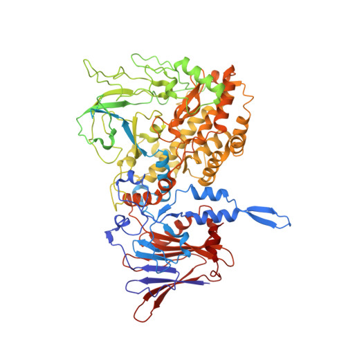
    Viruses sense environmental cues such as pH to engage in membrane interactions for cell entry during infection, but how nonenveloped viruses sense pH is largely undefined. Here, we report both high- and low-pH structures of bluetongue virus (BTV), which enters cells via a two-stage endosomal process. The receptor-binding protein VP2 possesses a zinc finger that may function to maintain VP2 in a metastable state and a conserved His866, which senses early-endosomal pH. The membrane-penetration protein VP5 has three domains: dagger, unfurling and anchoring. Notably, the β-meander motif of the anchoring domain contains a histidine cluster that can sense late-endosomal pH and also possesses four putative membrane-interaction elements. Exposing BTV to low pH detaches VP2 and dramatically refolds the dagger and unfurling domains of VP5. Our biochemical and structure-guided-mutagenesis studies support these coordinated pH-sensing mechanisms.


  • Organizational Affiliation
    • California NanoSystems Institute, University of California, Los Angeles (UCLA), Los Angeles, California, USA.

Macromolecule Content 

  • Total Structure Weight: 112.19 kDa 
  • Atom Count: 6,086 
  • Modeled Residue Count: 746 
  • Deposited Residue Count: 961 
  • Unique protein chains: 1

Macromolecules

Find similar proteins by:|  3D Structure
Entity ID: 1
MoleculeChains  Sequence LengthOrganismDetailsImage
Outer capsid protein VP2961Bluetongue virus 1Mutation(s): 0 
UniProt
Find proteins for B7U676 (Bluetongue virus 1)
Explore B7U676 
Go to UniProtKB:  B7U676
Entity Groups
Sequence Clusters30% Identity50% Identity70% Identity90% Identity95% Identity100% Identity
UniProt GroupB7U676
Sequence Annotations
Expand
Reference Sequence

Small Molecules

Ligands 1 Unique
IDChains Name / Formula / InChI Key2D Diagram3D Interactions
ZN

Query on ZN


Download:Ideal Coordinates CCD File
B [auth A]ZINC ION
Zn
PTFCDOFLOPIGGS-UHFFFAOYSA-N

Experimental Data & Validation

Experimental Data

  • Method: ELECTRON MICROSCOPY
  • Resolution: 3.30 Å
  • Aggregation State: PARTICLE 
  • Reconstruction Method: SINGLE PARTICLE 
EM Software:
TaskSoftware PackageVersion
RECONSTRUCTIONFREALIGN

Structure Validation

View Full Validation Report


Entry History 

Deposition Data

Revision History  (Full details and data files)

  • Version 1.0: 2015-12-09
    Type: Initial release
  • Version 1.1: 2015-12-23
    Changes: Database references
  • Version 1.2: 2016-01-20
    Changes: Database references
  • Version 1.3: 2018-07-18
    Changes: Data collection
  • Version 1.4: 2019-12-18
    Changes: Other
  • Version 1.5: 2024-02-21
    Changes: Data collection, Database references, Derived calculations, Refinement description