3J5M | pdb_00003j5m

Cryo-EM structure of the BG505 SOSIP.664 HIV-1 Env trimer with 3 PGV04 Fabs


Experimental Data Snapshot

  • Method: ELECTRON MICROSCOPY
  • Resolution: 5.80 Å
  • Aggregation State: PARTICLE 
  • Reconstruction Method: SINGLE PARTICLE 

Starting Models: experimental
View more details

wwPDB Validation 3D Report Full Report

Validation slider image for 3J5M

This is version 1.3 of the entry. See complete history

Literature

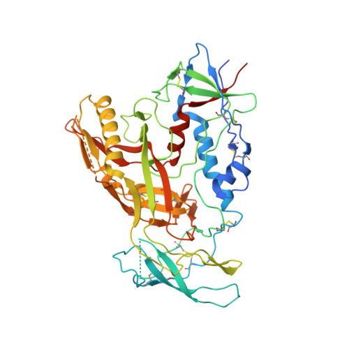
Cryo-EM structure of a fully glycosylated soluble cleaved HIV-1 envelope trimer.

Lyumkis, D.Julien, J.P.de Val, N.Cupo, A.Potter, C.S.Klasse, P.J.Burton, D.R.Sanders, R.W.Moore, J.P.Carragher, B.Wilson, I.A.Ward, A.B.

(2013) Science 342: 1484-1490

  • DOI: https://doi.org/10.1126/science.1245627
  • Primary Citation Related Structures: 
    3J5M

  • PubMed Abstract: 

    The HIV-1 envelope glycoprotein (Env) trimer contains the receptor binding sites and membrane fusion machinery that introduce the viral genome into the host cell. As the only target for broadly neutralizing antibodies (bnAbs), Env is a focus for rational vaccine design. We present a cryo-electron microscopy reconstruction and structural model of a cleaved, soluble Env trimer (termed BG505 SOSIP.664 gp140) in complex with a CD4 binding site (CD4bs) bnAb, PGV04, at 5.8 angstrom resolution. The structure reveals the spatial arrangement of Env components, including the V1/V2, V3, HR1, and HR2 domains, as well as shielding glycans. The structure also provides insights into trimer assembly, gp120-gp41 interactions, and the CD4bs epitope cluster for bnAbs, which covers a more extensive area and defines a more complex site of vulnerability than previously described.


  • Organizational Affiliation
    • National Resource for Automated Molecular Microscopy, The Scripps Research Institute, La Jolla, CA 92037, USA.

Macromolecule Content 

  • Total Structure Weight: 318.91 kDa 
  • Atom Count: 20,379 
  • Modeled Residue Count: 2,733 
  • Deposited Residue Count: 2,925 
  • Unique protein chains: 4

Macromolecules

Find similar proteins by:|  3D Structure
Entity ID: 1
MoleculeChains  Sequence LengthOrganismDetailsImage
BG505 SOSIP gp120
A, E, I
475Human immunodeficiency virus 1Mutation(s): 2 
Gene Names: env
UniProt
Find proteins for Q2N0S6 (Human immunodeficiency virus type 1)
Explore Q2N0S6 
Go to UniProtKB:  Q2N0S6
Entity Groups
Sequence Clusters30% Identity50% Identity70% Identity90% Identity95% Identity100% Identity
UniProt GroupQ2N0S6
Sequence Annotations
Expand
Reference Sequence
Find similar proteins by:|  3D Structure
Entity ID: 2
MoleculeChains  Sequence LengthOrganismDetailsImage
BG505 SOSIP gp41
B, F, J
64Human immunodeficiency virus 1Mutation(s): 0 
Gene Names: env
Find similar proteins by:|  3D Structure
Entity ID: 3
MoleculeChains  Sequence LengthOrganismDetailsImage
PGV04 light chain
C, G, K
208Homo sapiensMutation(s): 0 
Find similar proteins by:|  3D Structure
Entity ID: 4
MoleculeChains  Sequence LengthOrganismDetailsImage
PGV04 heavy chain
D, H, L
228Homo sapiensMutation(s): 0 

Experimental Data & Validation

Experimental Data

  • Method: ELECTRON MICROSCOPY
  • Resolution: 5.80 Å
  • Aggregation State: PARTICLE 
  • Reconstruction Method: SINGLE PARTICLE 
EM Software:
TaskSoftware PackageVersion
RECONSTRUCTIONFREALIGN
RECONSTRUCTIONXmipp

Structure Validation

View Full Validation Report


Entry History 

Deposition Data

Revision History  (Full details and data files)

  • Version 1.0: 2013-11-13
    Type: Initial release
  • Version 1.1: 2014-01-08
    Changes: Database references
  • Version 1.2: 2018-07-18
    Changes: Data collection
  • Version 1.3: 2024-11-27
    Changes: Data collection, Database references, Refinement description, Structure summary