3J2U | pdb_00003j2u

Kinesin-13 KLP10A HD in complex with CS-tubulin and a microtubule


Experimental Data Snapshot

  • Method: ELECTRON MICROSCOPY
  • Resolution: 10.8 Å
  • Aggregation State: FILAMENT 
  • Reconstruction Method: HELICAL 

Starting Model: experimental
View more details

wwPDB Validation 3D Report Full Report

Validation slider image for 3J2U

This is version 1.3 of the entry. See complete history

Literature

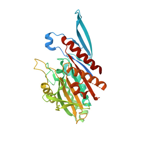
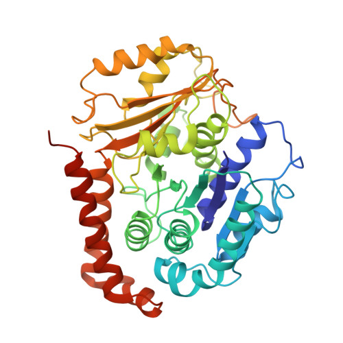
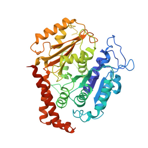
Structural model for tubulin recognition and deformation by Kinesin-13 microtubule depolymerases.

Asenjo, A.B.Chatterjee, C.Tan, D.Depaoli, V.Rice, W.J.Diaz-Avalos, R.Silvestry, M.Sosa, H.

(2013) Cell Rep 3: 759-768

  • DOI: https://doi.org/10.1016/j.celrep.2013.01.030
  • Primary Citation Related Structures: 
    3J2U

  • PubMed Abstract: 

    To elucidate the structural basis of the mechanism of microtubule depolymerization by kinesin-13s, we analyzed complexes of tubulin and the Drosophila melanogaster kinesin-13 KLP10A by electron microscopy (EM) and fluorescence polarization microscopy. We report a nanometer-resolution (1.1 nm) cryo-EM three-dimensional structure of the KLP10A head domain (KLP10AHD) bound to curved tubulin. We found that binding of KLP10AHD induces a distinct tubulin configuration with displacement (shear) between tubulin subunits in addition to curvature. In this configuration, the kinesin-binding site differs from that in straight tubulin, providing an explanation for the distinct interaction modes of kinesin-13s with the microtubule lattice or its ends. The KLP10AHD-tubulin interface comprises three areas of interaction, suggesting a crossbow-type tubulin-bending mechanism. These areas include the kinesin-13 family conserved KVD residues, and as predicted from the crossbow model, mutating these residues changes the orientation and mobility of KLP10AHDs interacting with the microtubule.


  • Organizational Affiliation
    • Department of Physiology and Biophysics, Albert Einstein College of Medicine, Bronx, NY 10461, USA.

Macromolecule Content 

  • Total Structure Weight: 241.84 kDa 
  • Atom Count: 15,575 
  • Modeled Residue Count: 1,985 
  • Deposited Residue Count: 2,166 
  • Unique protein chains: 3

Macromolecules

Find similar proteins by:|  3D Structure
Entity ID: 1
MoleculeChains  Sequence LengthOrganismDetailsImage
Kinesin-like protein Klp10AA [auth K]374Drosophila melanogasterMutation(s): 0 
Gene Names: Klp10ACG1453
UniProt
Find proteins for Q960Z0 (Drosophila melanogaster)
Explore Q960Z0 
Go to UniProtKB:  Q960Z0
Entity Groups
Sequence Clusters30% Identity50% Identity70% Identity90% Identity95% Identity100% Identity
UniProt GroupQ960Z0
Sequence Annotations
Expand
Reference Sequence
Find similar proteins by:|  3D Structure
Entity ID: 2
MoleculeChains  Sequence LengthOrganismDetailsImage
Tubulin alpha-1A chainB [auth A],
D [auth C]
451Bos taurusMutation(s): 0 
EC: 3.6.5
UniProt
Find proteins for P81947 (Bos taurus)
Explore P81947 
Go to UniProtKB:  P81947
Entity Groups
Sequence Clusters30% Identity50% Identity70% Identity90% Identity95% Identity100% Identity
UniProt GroupP81947
Sequence Annotations
Expand
Reference Sequence
Find similar proteins by:|  3D Structure
Entity ID: 3
MoleculeChains  Sequence LengthOrganismDetailsImage
Tubulin beta-2B chainC [auth B],
E [auth D]
445Bos taurusMutation(s): 0 
UniProt
Find proteins for Q6B856 (Bos taurus)
Explore Q6B856 
Go to UniProtKB:  Q6B856
Entity Groups
Sequence Clusters30% Identity50% Identity70% Identity90% Identity95% Identity100% Identity
UniProt GroupQ6B856
Sequence Annotations
Expand
Reference Sequence

Experimental Data & Validation

Experimental Data

  • Method: ELECTRON MICROSCOPY
  • Resolution: 10.8 Å
  • Aggregation State: FILAMENT 
  • Reconstruction Method: HELICAL 
EM Software:
TaskSoftware PackageVersion
RECONSTRUCTIONCustom
RECONSTRUCTIONIHRSR
RECONSTRUCTIONSPIDER

Structure Validation

View Full Validation Report


Entry History 

Deposition Data

Revision History  (Full details and data files)

  • Version 1.0: 2013-03-06
    Type: Initial release
  • Version 1.1: 2013-04-10
    Changes: Database references
  • Version 1.2: 2018-07-18
    Changes: Author supporting evidence, Data collection
  • Version 1.3: 2024-02-21
    Changes: Data collection, Database references, Derived calculations, Refinement description