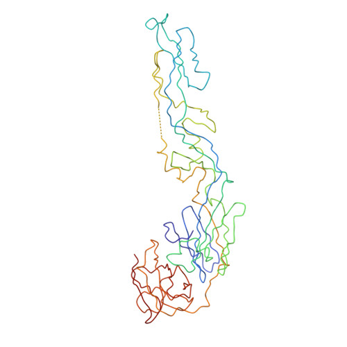
3J0B

cryo-EM reconstruction of West Nile virus


Experimental Data Snapshot

  • Method: ELECTRON MICROSCOPY
  • Resolution: 10.3 Å
  • Aggregation State: PARTICLE 
  • Reconstruction Method: SINGLE PARTICLE 

Starting Model: experimental
View more details

wwPDB Validation   3D Report Full Report


This is version 1.4 of the entry. See complete history


Literature

Membrane curvature in flaviviruses.

Zhang, W.Kaufmann, B.Chipman, P.R.Kuhn, R.J.Rossmann, M.G.

(2013) J Struct Biol 183: 86-94

  • DOI: https://doi.org/10.1016/j.jsb.2013.04.005
  • Primary Citation of Related Structures:  
    3J0B

  • PubMed Abstract: 

    Coordinated interplay between membrane proteins and the lipid bilayer is required for such processes as transporter function and the entrance of enveloped viruses into host cells. In this study, three-dimensional cryo-electron microscopy density maps of mature and immature flaviviruses were analyzed to assess the curvature of the membrane leaflets and its relation to membrane-bound viral glycoproteins. The overall morphology of the viral membrane is determined by the icosahedral scaffold composed of envelope (E) and membrane (M) proteins through interaction of the proteins' stem-anchor regions with the membrane. In localized regions, small membrane areas exhibit convex, concave, flat or saddle-shaped surfaces that are constrained by the specific protein organization within each membrane leaflet. These results suggest that the organization of membrane proteins in small enveloped viruses mediate the formation of membrane curvature.


  • Organizational Affiliation

    Department of Biological Sciences, Purdue University, West Lafayette, IN 47907, USA. zhangwei@umn.edu


Macromolecules
Find similar proteins by:  (by identity cutoff)  |  3D Structure
Entity ID: 1
MoleculeChains Sequence LengthOrganismDetailsImage
envelope glycoprotein E
A, B, C
400West Nile virusMutation(s): 0 
UniProt
Find proteins for Q9Q6P4 (West Nile virus (strain NY-99))
Go to UniProtKB:  Q9Q6P4
Sequence Annotations
Expand
  • Reference Sequence
Experimental Data & Validation

Experimental Data

  • Method: ELECTRON MICROSCOPY
  • Resolution: 10.3 Å
  • Aggregation State: PARTICLE 
  • Reconstruction Method: SINGLE PARTICLE 
EM Software:
TaskSoftware PackageVersion
RECONSTRUCTIONAuto3DEM

Structure Validation

View Full Validation Report


Entry History 

Deposition Data

Revision History  (Full details and data files)

  • Version 1.0: 2012-12-19
    Type: Initial release
  • Version 1.1: 2013-05-01
    Changes: Database references
  • Version 1.2: 2013-07-24
    Changes: Database references
  • Version 1.3: 2018-07-18
    Changes: Data collection
  • Version 1.4: 2024-02-21
    Changes: Data collection, Database references, Derived calculations, Refinement description