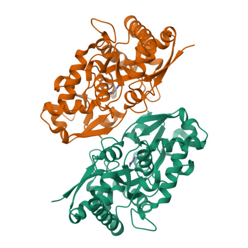
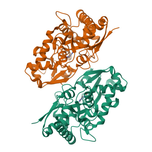
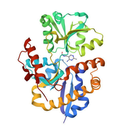
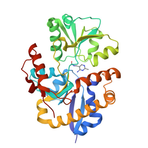
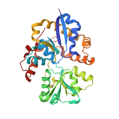
3IX1

Periplasmic N-formyl-4-amino-5-aminomethyl-2-methylpyrimidine binding protein from Bacillus halodurans


Experimental Data Snapshot

  • Method: X-RAY DIFFRACTION
  • Resolution: 2.40 Å
  • R-Value Free: 0.264 
  • R-Value Work: 0.203 
  • R-Value Observed: 0.203 

wwPDB Validation   3D Report Full Report


Ligand Structure Quality Assessment 


This is version 1.3 of the entry. See complete history


Literature

HMP Binding Protein ThiY and HMP-P Synthase THI5 Are Structural Homologues.

Bale, S.Rajashankar, K.R.Perry, K.Begley, T.P.Ealick, S.E.

(2010) Biochemistry 49: 8929-8936

  • DOI: https://doi.org/10.1021/bi101209t
  • Primary Citation of Related Structures:  
    3IX1

  • PubMed Abstract: 

    The ATP-binding cassette transporter system ThiXYZ transports N-formyl-4-amino-5-(aminomethyl)-2-methylpyrimidine (FAMP), a thiamin salvage pathway intermediate, into cells. FAMP is then converted to 4-amino-5-(hydroxymethyl)-2-methylpyrimidine (HMP) and recycled into the thiamin biosynthetic pathway. ThiY is the periplasmic substrate binding protein of the ThiXYZ system and delivers the substrate FAMP to the transmembrane domain. We report the crystal structure of Bacillus halodurans ThiY with FAMP bound at 2.4 Å resolution determined by single-wavelength anomalous diffraction phasing. The crystal structure reveals that ThiY belongs to the group II periplasmic binding protein family. The closest structural homologues of ThiY are periplasmic binding proteins involved in alkanesulfonate/nitrate and bicarbonate transport. ThiY is also structurally homologous to thiamin binding protein (TbpA) and to thiaminase-I. THI5 is responsible for the synthesis of 4-amino-5-(hydroxymethyl)-2-methylpyrimidine phosphate in the thiamin biosynthetic pathway of eukaryotes and is approximately 25% identical in sequence with ThiY. A homology model of Saccharomyces cerevisiae THI5 was generated on the basis of the structure of ThiY. Many features of the thiamin pyrimidine binding site are shared between ThiY and THI5, suggesting a common ancestor.


  • Organizational Affiliation

    Department of Chemistry and Chemical Biology, Cornell University, Ithaca, NY 14853, USA.


Macromolecules
Find similar proteins by:  (by identity cutoff)  |  3D Structure
Entity ID: 1
MoleculeChains Sequence LengthOrganismDetailsImage
N-formyl-4-amino-5-aminomethyl-2-methylpyrimidine binding protein
A, B
302Halalkalibacterium halodurans C-125Mutation(s): 0 
Gene Names: BH2682
UniProt
Find proteins for Q9K9G5 (Halalkalibacterium halodurans (strain ATCC BAA-125 / DSM 18197 / FERM 7344 / JCM 9153 / C-125))
Explore Q9K9G5 
Go to UniProtKB:  Q9K9G5
Entity Groups  
Sequence Clusters30% Identity50% Identity70% Identity90% Identity95% Identity100% Identity
UniProt GroupQ9K9G5
Sequence Annotations
Expand
  • Reference Sequence
Small Molecules
Ligands 1 Unique
IDChains Name / Formula / InChI Key2D Diagram3D Interactions
NFM
Query on NFM

Download Ideal Coordinates CCD File 
C [auth A],
D [auth B]
N-[(4-amino-2-methylpyrimidin-5-yl)methyl]formamide
C7 H10 N4 O
PVWNFAGYFUUDRC-UHFFFAOYSA-N
Experimental Data & Validation

Experimental Data

  • Method: X-RAY DIFFRACTION
  • Resolution: 2.40 Å
  • R-Value Free: 0.264 
  • R-Value Work: 0.203 
  • R-Value Observed: 0.203 
  • Space Group: P 21 21 21
Unit Cell:
Length ( Å )Angle ( ˚ )
a = 59.918α = 90
b = 96.953β = 90
c = 104.888γ = 90
Software Package:
Software NamePurpose
DENZOdata reduction
SCALEPACKdata scaling
CNSrefinement
PDB_EXTRACTdata extraction
ADSCdata collection
HKL-2000data reduction
HKL-2000data scaling
SHELXDphasing

Structure Validation

View Full Validation Report


Ligand Structure Quality Assessment 


Entry History 

Deposition Data

Revision History  (Full details and data files)

  • Version 1.0: 2010-10-13
    Type: Initial release
  • Version 1.1: 2011-07-13
    Changes: Version format compliance
  • Version 1.2: 2017-11-01
    Changes: Refinement description
  • Version 1.3: 2024-02-21
    Changes: Data collection, Database references, Derived calculations