3IWY | pdb_00003iwy

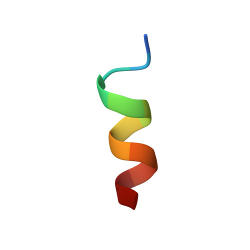
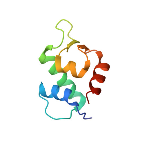
Crystal structure of human MDM2 complexed with D-peptide (12 residues)


Experimental Data Snapshot

  • Method: X-RAY DIFFRACTION
  • Resolution: 1.93 Å
  • R-Value Free: 
    0.259 (Depositor), 0.257 (DCC) 
  • R-Value Work: 
    0.210 (Depositor), 0.211 (DCC) 
  • R-Value Observed: 
    0.213 (Depositor) 

Starting Model: experimental
View more details

wwPDB Validation 3D Report Full Report

Validation slider image for 3IWY

This is version 1.5 of the entry. See complete history. 

Literature

D-peptide inhibitors of the p53-MDM2 interaction for targeted molecular therapy of malignant neoplasms.

Liu, M., Li, C., Pazgier, M., Li, C., Mao, Y., Lv, Y., Gu, B., Wei, G., Yuan, W., Zhan, C., Lu, W.Y., Lu, W.

(2010) Proc Natl Acad Sci U S A 398: 200-213

  • DOI: https://doi.org/10.1073/pnas.1008930107
  • Primary Citation Related Structures: 
    3IWY

  • PubMed Abstract: 

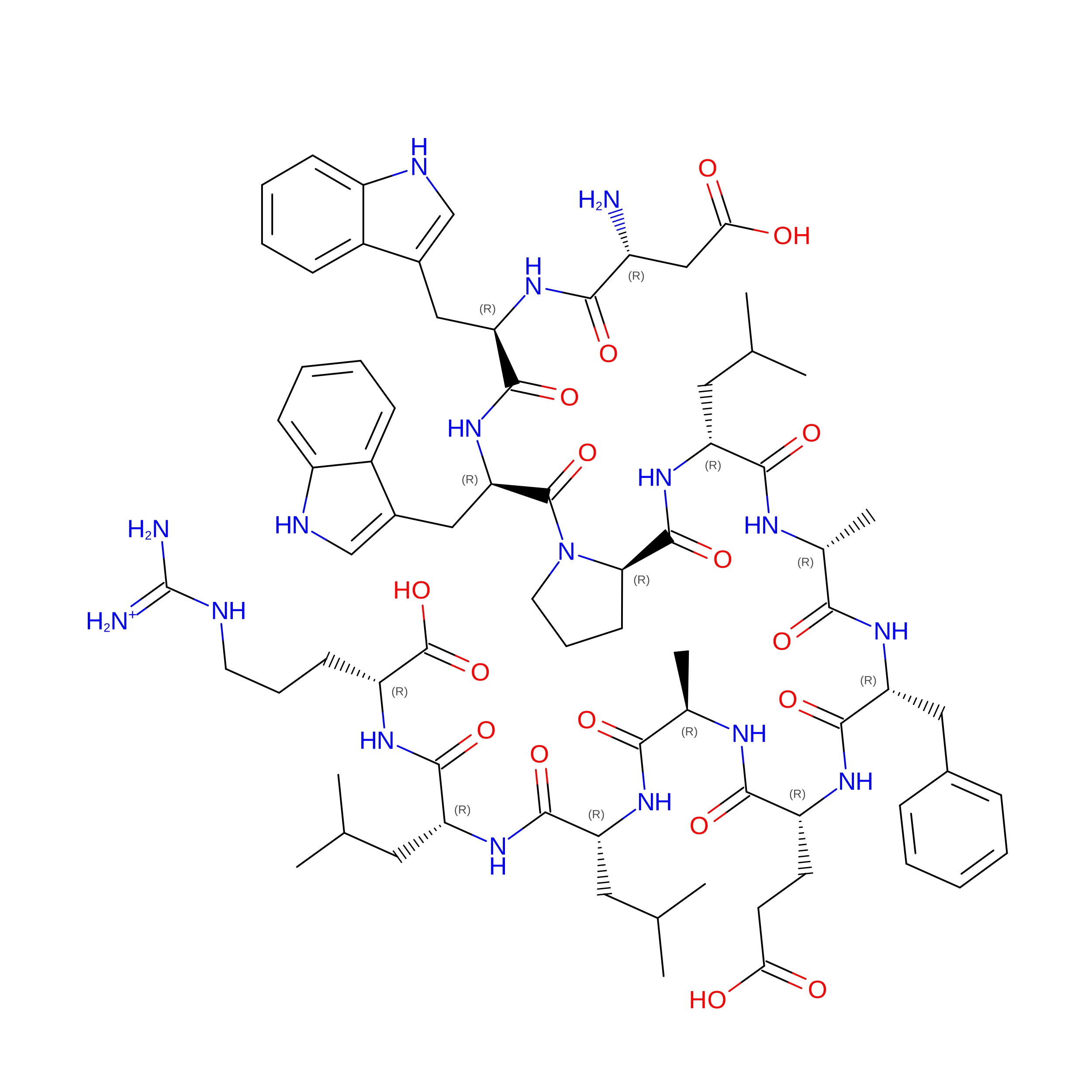
    The oncoproteins MDM2 and MDMX negatively regulate the activity and stability of the tumor suppressor protein p53, conferring tumor development and survival. Antagonists targeting the p53-binding domains of MDM2 and MDMX kill tumor cells both in vitro and in vivo by reactivating the p53 pathway, promising a class of antitumor agents for cancer therapy. Aided by native chemical ligation and mirror image phage display, we recently identified a D-peptide inhibitor of the p53-MDM2 interaction termed (D)PMI-alpha (TNWYANLEKLLR) that competes with p53 for MDM2 binding at an affinity of 219 nM. Increased selection stringency resulted in a distinct D-peptide inhibitor termed (D)PMI-gamma (DWWPLAFEALLR) that binds MDM2 at an affinity of 53 nM. Structural studies coupled with mutational analysis verified the mode of action of these D-peptides as MDM2-dependent p53 activators. Despite being resistant to proteolysis, both (D)PMI-alpha and (D)PMI-gamma failed to actively traverse the cell membrane and, when conjugated to a cationic cell-penetrating peptide, were indiscriminately cytotoxic independently of p53 status. When encapsulated in liposomes decorated with an integrin-targeting cyclic-RGD peptide, however, (D)PMI-alpha exerted potent p53-dependent growth inhibitory activity against human glioblastoma in cell cultures and nude mouse xenograft models. Our findings validate D-peptide antagonists of MDM2 as a class of p53 activators for targeted molecular therapy of malignant neoplasms harboring WT p53 and elevated levels of MDM2.


  • Organizational Affiliation: 
    • Institute of Human Virology and Department of Biochemistry and Molecular Biology, University of Maryland School of Medicine, Baltimore, MD 21201, USA.

Macromolecule Content 

  • Total Structure Weight: 23.13 kDa 
  • Atom Count: 1,701 
  • Modeled Residue Count: 189 
  • Deposited Residue Count: 194 
  • Unique protein chains: 2

Macromolecules

Find similar proteins by:  Sequence   |   3D Structure  
Entity ID: 1
MoleculeChains  Sequence LengthOrganismDetailsImage
D-peptide inhibitorA [auth B],
B [auth D]
12N/AMutation(s): 0 
Entity Groups
Sequence Clusters30% Identity50% Identity70% Identity90% Identity95% Identity100% Identity
Sequence Annotations
Expand
Reference Sequence
Find similar proteins by:|  3D Structure
Entity ID: 2
MoleculeChains  Sequence LengthOrganismDetailsImage
E3 ubiquitin-protein ligase Mdm2C [auth A],
D [auth C]
85N/AMutation(s): 0 
EC: 6.3.2 (PDB Primary Data), 2.3.2.27 (UniProt)
UniProt & NIH Common Fund Data Resources
Find proteins for Q00987 (Homo sapiens)
Explore Q00987 
Go to UniProtKB:  Q00987
PHAROS:  Q00987
GTEx:  ENSG00000135679 
Entity Groups
Sequence Clusters30% Identity50% Identity70% Identity90% Identity95% Identity100% Identity
UniProt GroupQ00987
Sequence Annotations
Expand
Reference Sequence

Small Molecules

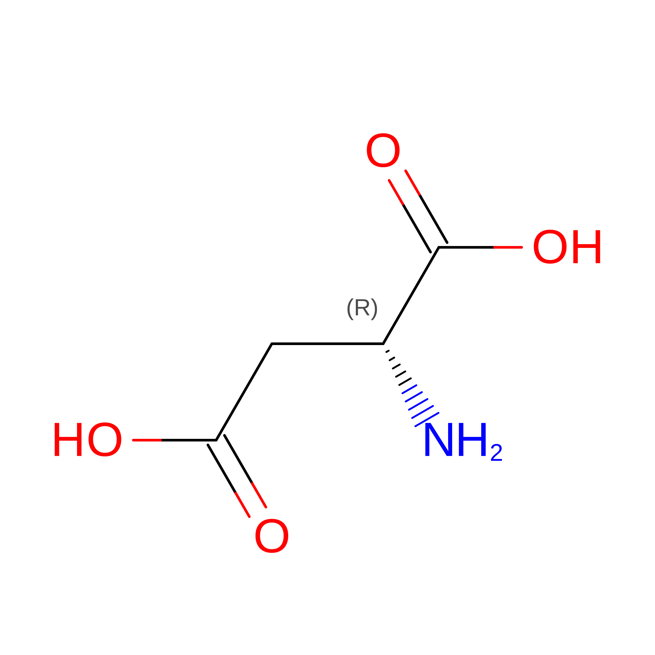
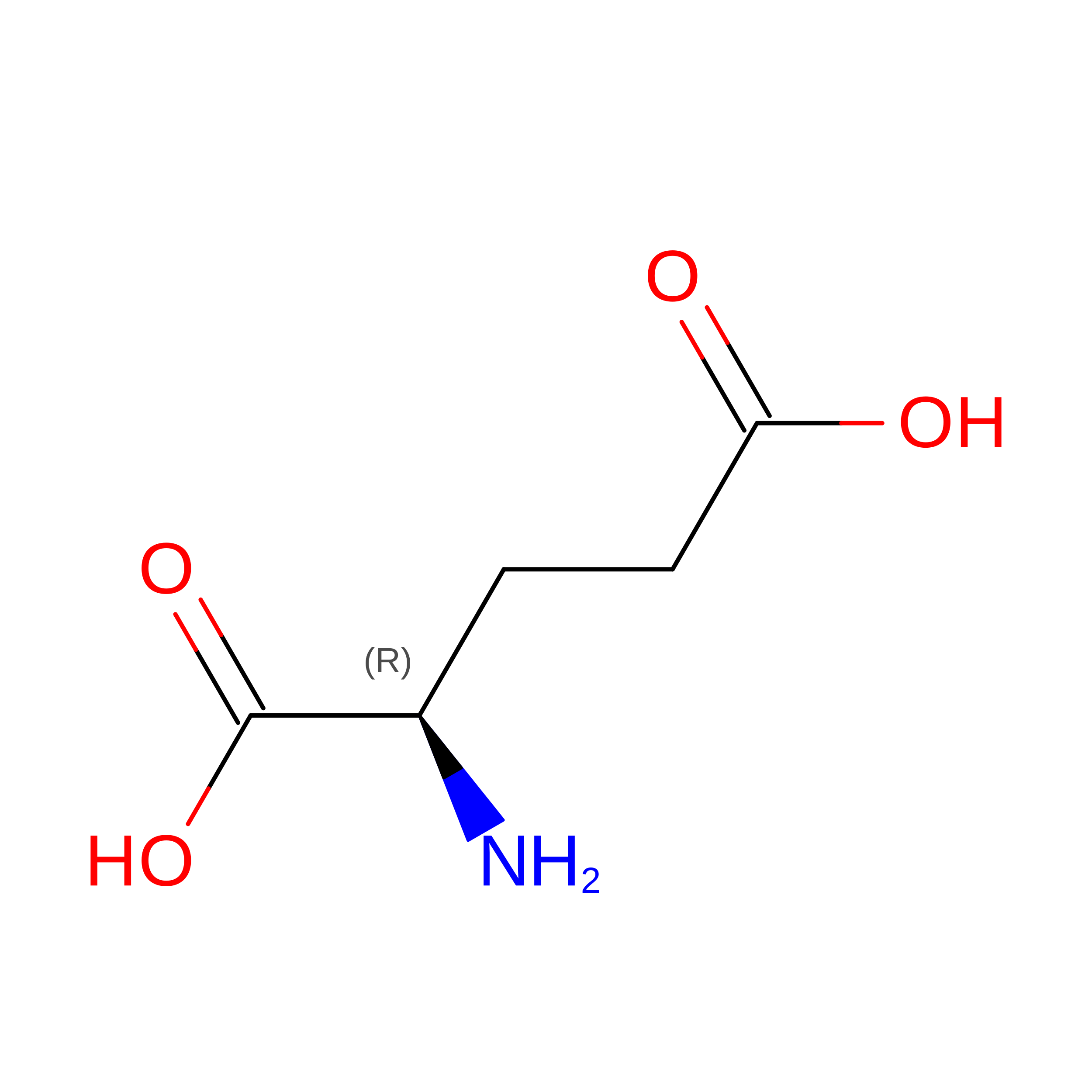
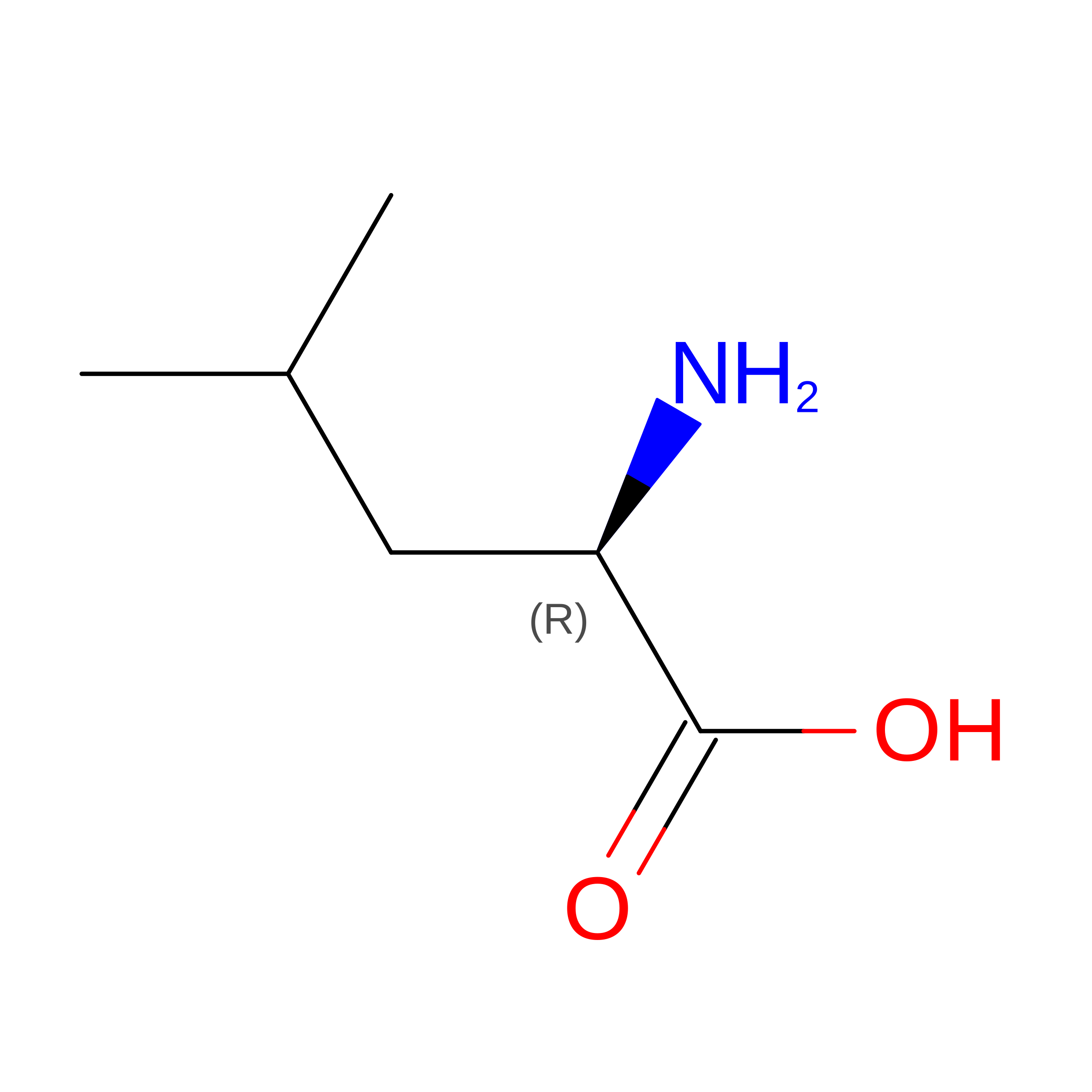
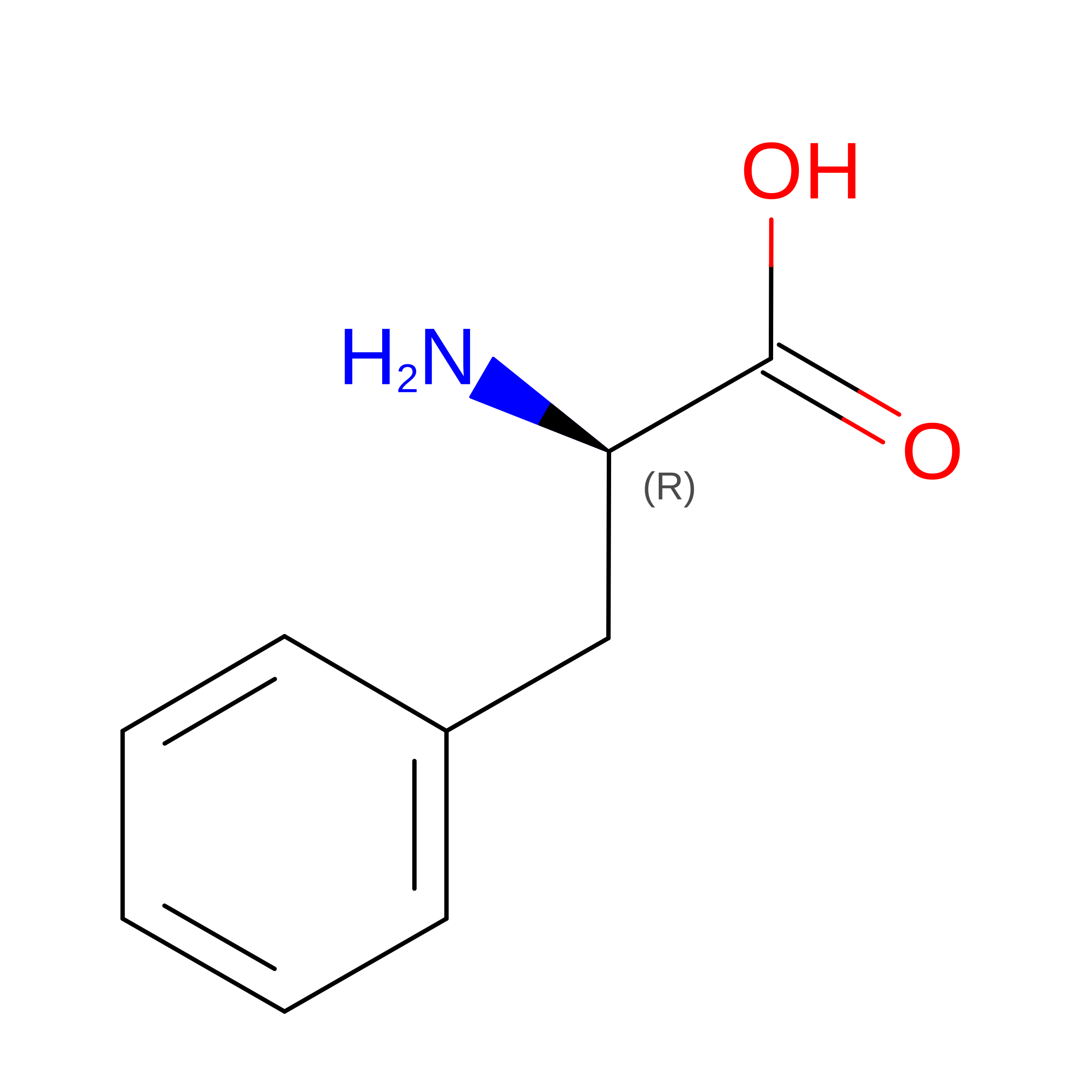
Modified Residues  8 Unique
IDChains TypeFormula2D DiagramParent
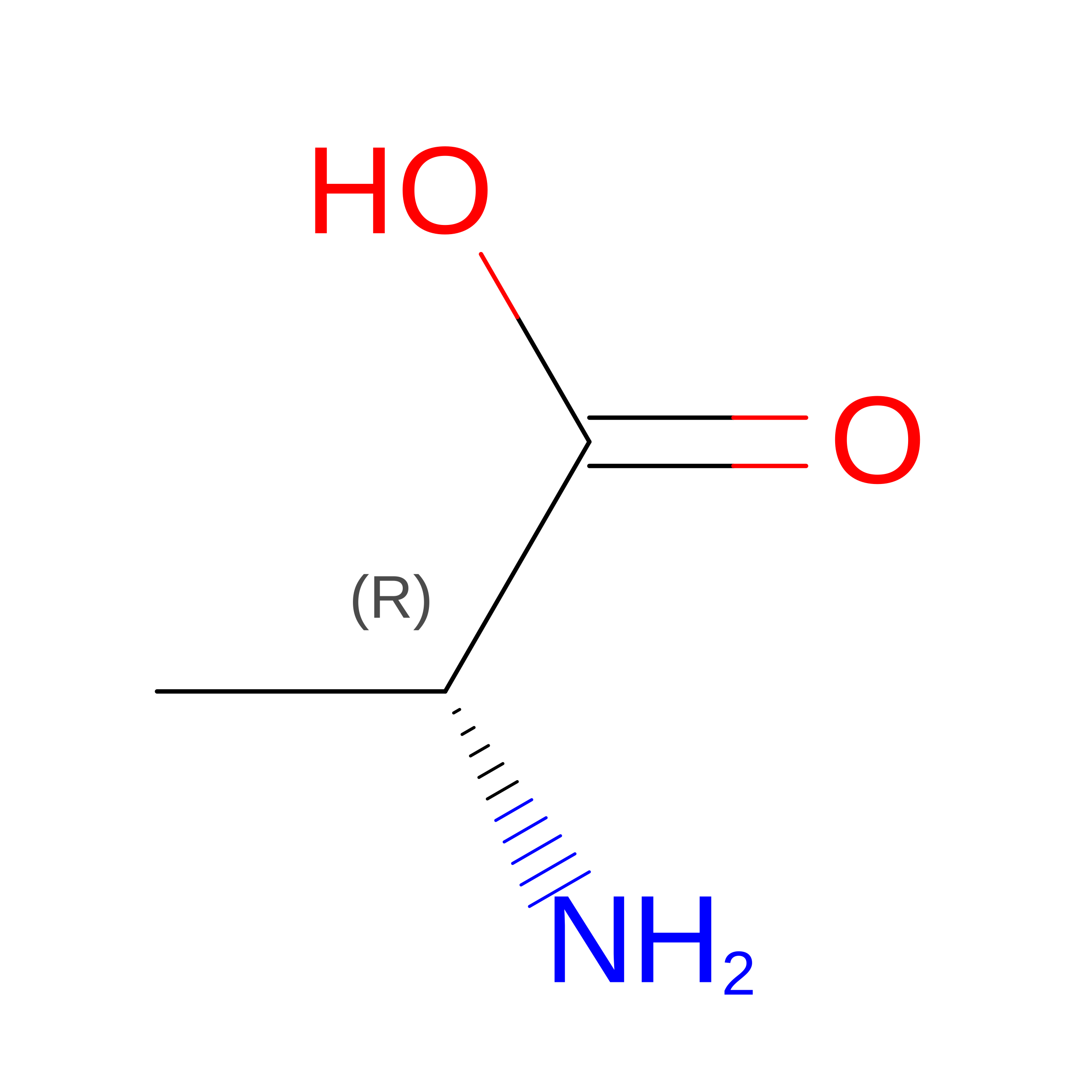
DAL
Query on DAL
A [auth B],
B [auth D]
D-PEPTIDE LINKINGC3 H7 N O2

--

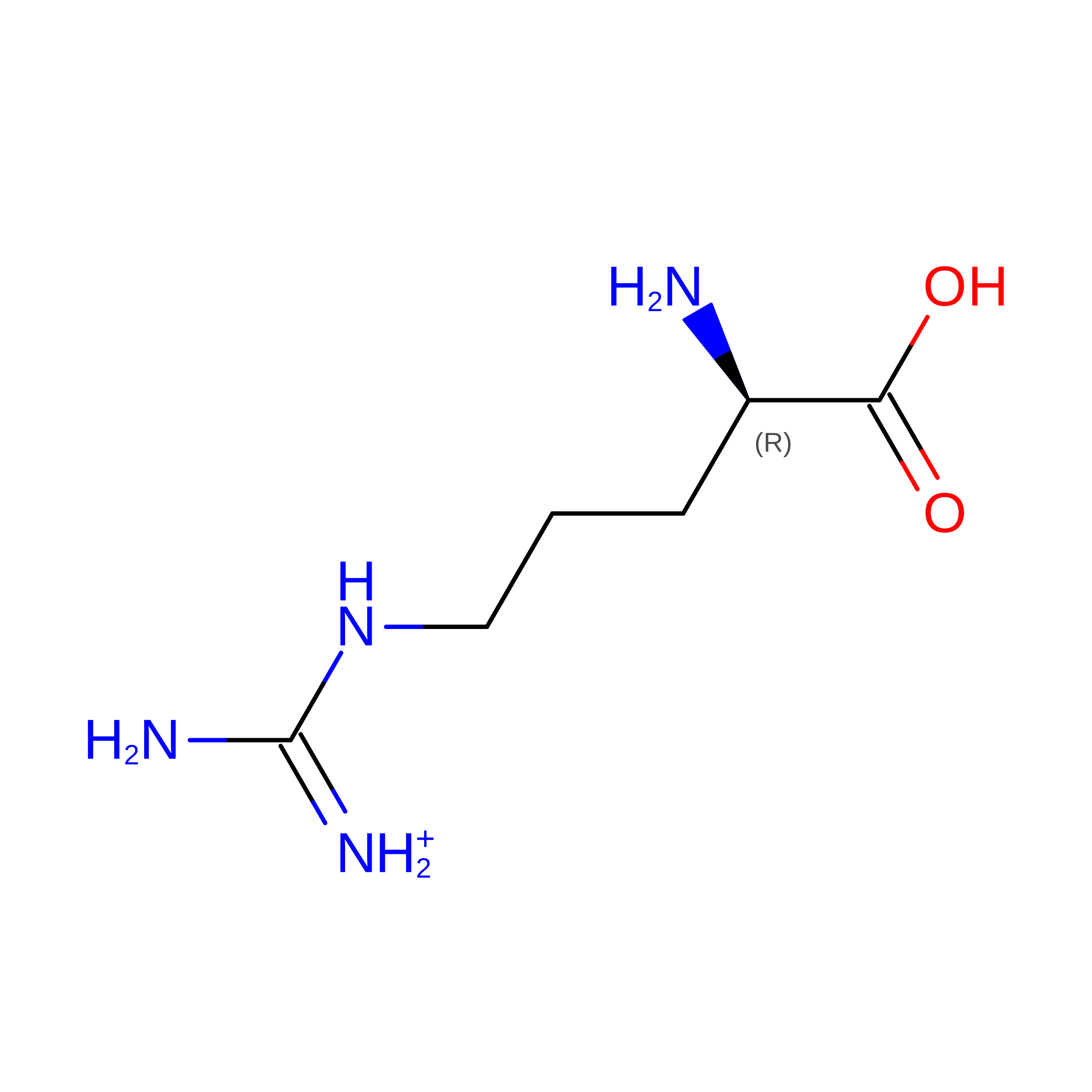
DAR
Query on DAR
A [auth B],
B [auth D]
D-PEPTIDE LINKINGC6 H15 N4 O2

--

DAS
Query on DAS
A [auth B],
B [auth D]
D-PEPTIDE LINKINGC4 H7 N O4

--

DGL
Query on DGL
A [auth B],
B [auth D]
D-PEPTIDE LINKINGC5 H9 N O4

--

DLE
Query on DLE
A [auth B],
B [auth D]
D-PEPTIDE LINKINGC6 H13 N O2

--

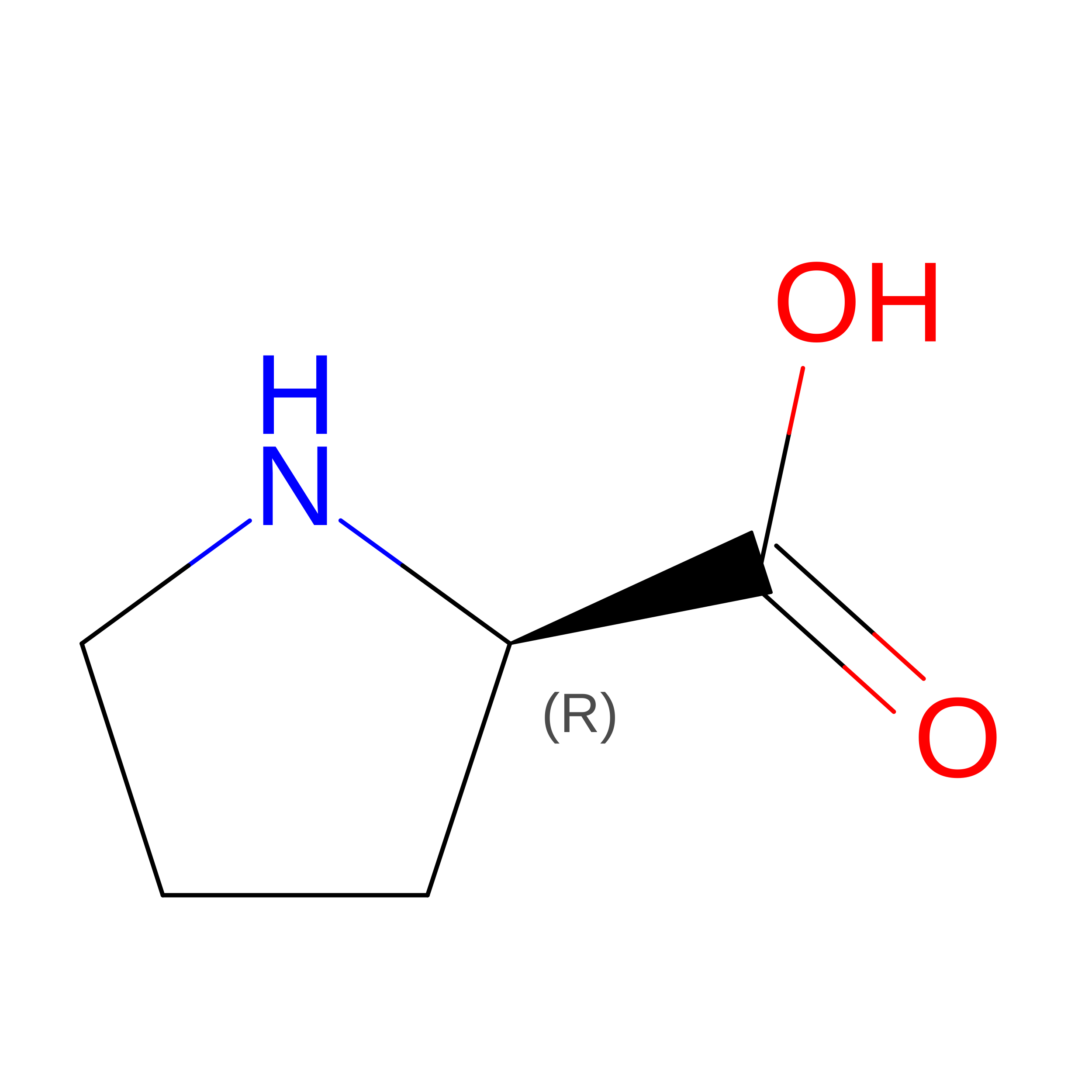
DPN
Query on DPN
A [auth B],
B [auth D]
D-PEPTIDE LINKINGC9 H11 N O2

--

DPR
Query on DPR
A [auth B],
B [auth D]
D-PEPTIDE LINKINGC5 H9 N O2

--

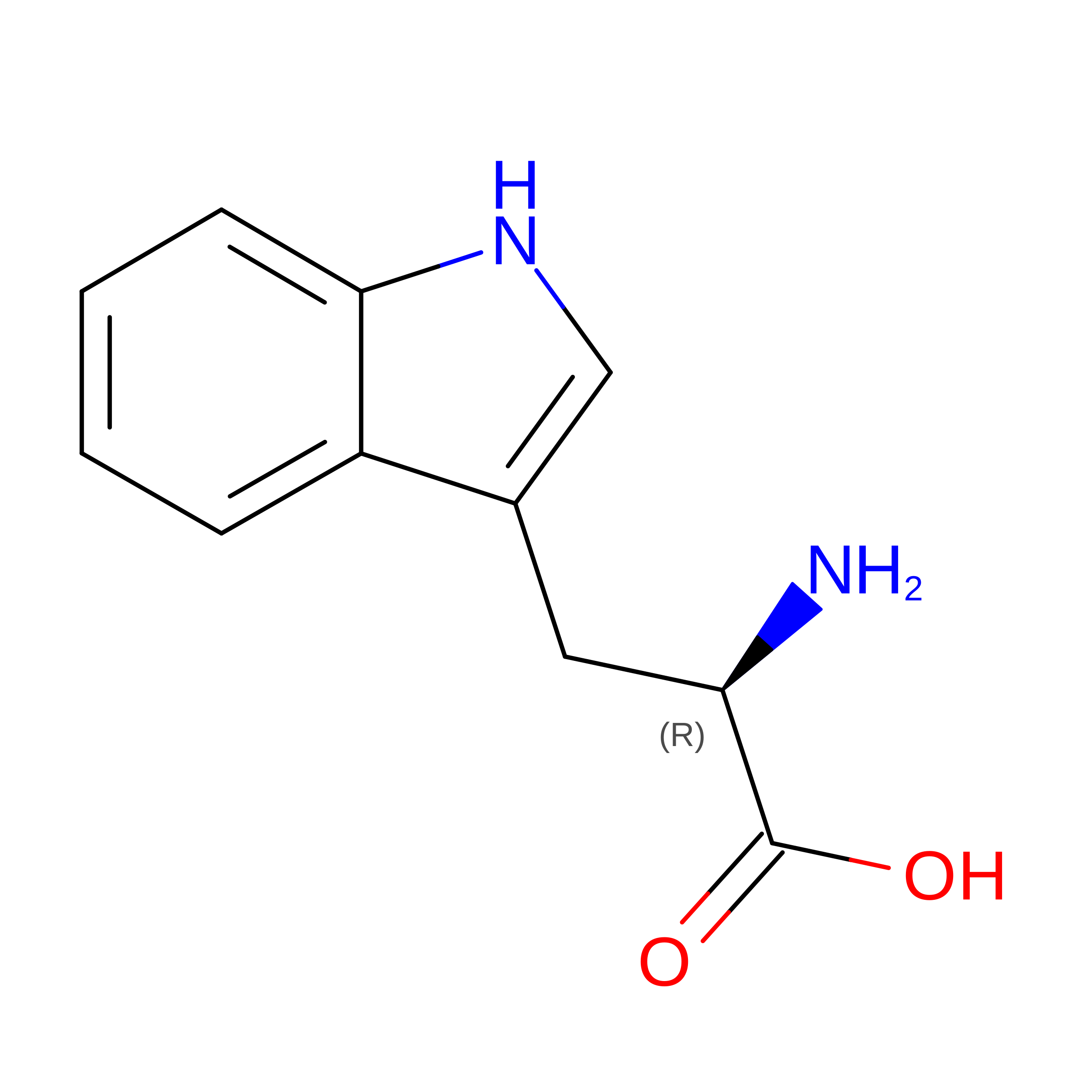
DTR
Query on DTR
A [auth B],
B [auth D]
D-PEPTIDE LINKINGC11 H12 N2 O2

--

Biologically Interesting Molecules (External Reference) 

1 Unique

Experimental Data & Validation

Experimental Data

  • Method: X-RAY DIFFRACTION
  • Resolution: 1.93 Å
  • R-Value Free:  0.259 (Depositor), 0.257 (DCC) 
  • R-Value Work:  0.210 (Depositor), 0.211 (DCC) 
  • R-Value Observed: 0.213 (Depositor) 
Space Group: P 1 21 1
Unit Cell:
Length ( Å )Angle ( ˚ )
a = 33.569α = 90
b = 46.503β = 92.35
c = 49.353γ = 90
Software Package:
Software NamePurpose
HKL-2000data collection
PHASERphasing
REFMACrefinement
DENZOdata reduction
SCALEPACKdata scaling

Structure Validation

View Full Validation Report


Entry History 

Deposition Data

Revision History  (Full details and data files)

  • Version 1.0: 2010-04-21
    Type: Initial release
  • Version 1.1: 2011-07-13
    Changes: Advisory, Refinement description, Version format compliance
  • Version 1.2: 2012-12-12
    Changes: Other
  • Version 1.3: 2023-09-06
    Changes: Data collection, Database references, Derived calculations, Refinement description
  • Version 1.4: 2023-11-22
    Changes: Data collection
  • Version 1.5: 2024-11-20
    Changes: Structure summary