3IVZ | pdb_00003ivz

Crystal structure of hyperthermophilic nitrilase


Experimental Data Snapshot

  • Method: X-RAY DIFFRACTION
  • Resolution: 1.57 Å
  • R-Value Free: 
    0.176 (Depositor), 0.172 (DCC) 
  • R-Value Work: 
    0.131 (Depositor), 0.132 (DCC) 
  • R-Value Observed: 
    0.132 (Depositor) 

Starting Model: experimental
View more details

wwPDB Validation 3D Report Full Report

Validation slider image for 3IVZ

This is version 1.2 of the entry. See complete history

Literature

Crystallographic analysis of a thermoactive nitrilase.

Raczynska, J.E.Vorgias, C.E.Antranikian, G.Rypniewski, W.

(2010) J Struct Biol 173: 294-302

  • DOI: https://doi.org/10.1016/j.jsb.2010.11.017
  • Primary Citation Related Structures: 
    3IVZ, 3IW3, 3KI8, 3KLC

  • PubMed Abstract: 

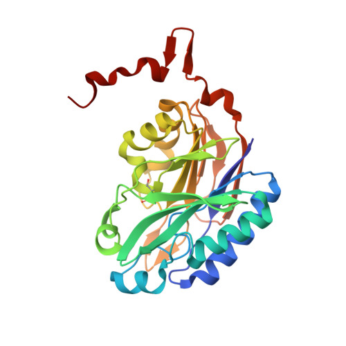
    The nitrilase superfamily is a large and diverse superfamily of enzymes that catalyse the cleavage of various types of carbon-nitrogen bonds using a Cys-Glu-Lys catalytic triad. Thermoactive nitrilase from Pyrococcus abyssi (PaNit) hydrolyses small aliphatic nitriles like fumaro- and malononitryl. Yet, the biological role of this enzyme is unknown. We have analysed several crystal structures of PaNit: without ligands, with an acetate ion bound in the active site and with a bromide ion in the active site. In addition, docking calculations have been performed for fumaro- and malononitriles. The structures provide a proof for specific binding of the carboxylate ion and a general affinity for negatively changed ligands. The role of residues in the active site is considered and an enzymatic reaction mechanism is proposed in which Cys146 acts as the nucleophile, Glu42 as the general base, Lys113/Glu42 as the general acid, WatA as the hydrolytic water and Nζ_Lys113 and N_Phe147 form the oxyanion hole.


  • Organizational Affiliation
    • Institute of Bioorganic Chemistry, Polish Academy of Sciences, Poznan, Poland.

Macromolecule Content 

  • Total Structure Weight: 59.57 kDa 
  • Atom Count: 4,314 
  • Modeled Residue Count: 522 
  • Deposited Residue Count: 524 
  • Unique protein chains: 1

Macromolecules

Find similar proteins by:|  3D Structure
Entity ID: 1
MoleculeChains  Sequence LengthOrganismDetailsImage
Nitrilase
A, B
262Pyrococcus abyssi GE5Mutation(s): 0 
Gene Names: nit-30PAB1449PYRAB13990
EC: 3.5.1.6 (PDB Primary Data), 3.5.5.1 (UniProt)
UniProt
Find proteins for Q9UYV8 (Pyrococcus abyssi (strain GE5 / Orsay))
Explore Q9UYV8 
Go to UniProtKB:  Q9UYV8
Entity Groups
Sequence Clusters30% Identity50% Identity70% Identity90% Identity95% Identity100% Identity
UniProt GroupQ9UYV8
Sequence Annotations
Expand
Reference Sequence

Small Molecules

Ligands 1 Unique
IDChains Name / Formula / InChI Key2D Diagram3D Interactions
MG

Query on MG


Download:Ideal Coordinates CCD File
C [auth A]MAGNESIUM ION
Mg
JLVVSXFLKOJNIY-UHFFFAOYSA-N
Modified Residues  1 Unique
IDChains TypeFormula2D DiagramParent
CSX
Query on CSX
A, B
L-PEPTIDE LINKINGC3 H7 N O3 SCYS

Experimental Data & Validation

Experimental Data

  • Method: X-RAY DIFFRACTION
  • Resolution: 1.57 Å
  • R-Value Free:  0.176 (Depositor), 0.172 (DCC) 
  • R-Value Work:  0.131 (Depositor), 0.132 (DCC) 
  • R-Value Observed: 0.132 (Depositor) 
Space Group: P 41
Unit Cell:
Length ( Å )Angle ( ˚ )
a = 59.174α = 90
b = 59.174β = 90
c = 127.22γ = 90
Software Package:
Software NamePurpose
ADSCdata collection
PHASERphasing
REFMACrefinement
DENZOdata reduction
SCALEPACKdata scaling

Structure Validation

View Full Validation Report


Entry History 

Deposition Data

Revision History  (Full details and data files)

  • Version 1.0: 2010-09-22
    Type: Initial release
  • Version 1.1: 2011-07-13
    Changes: Source and taxonomy, Version format compliance
  • Version 1.2: 2023-09-06
    Changes: Data collection, Database references, Derived calculations, Refinement description