3IUJ | pdb_00003iuj

apPEP_WT2 opened state


Experimental Data Snapshot

  • Method: X-RAY DIFFRACTION
  • Resolution: 1.80 Å
  • R-Value Free: 
    0.218 (Depositor), 0.194 (DCC) 
  • R-Value Work: 
    0.181 (Depositor), 0.192 (DCC) 
  • R-Value Observed: 
    0.181 (Depositor) 

wwPDB Validation 3D Report Full Report

Validation slider image for 3IUJ

This is version 1.3 of the entry. See complete history

Literature

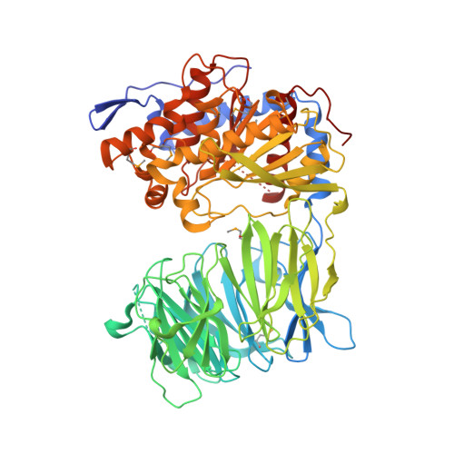
Induced-fit mechanism for prolyl endopeptidase

Li, M.Chen, C.Davies, D.R.Chiu, T.K.

(2010) J Biological Chem 285: 21487-21495

  • DOI: https://doi.org/10.1074/jbc.M109.092692
  • Primary Citation Related Structures: 
    3IUJ, 3IUL, 3IUM, 3IUN, 3IUQ, 3IUR, 3IVM

  • PubMed Abstract: 

    Prolyl peptidases cleave proteins at proline residues and are of importance for cancer, neurological function, and type II diabetes. Prolyl endopeptidase (PEP) cleaves neuropeptides and is a drug target for neuropsychiatric diseases such as post-traumatic stress disorder, depression, and schizophrenia. Previous structural analyses showing little differences between native and substrate-bound structures have suggested a lock-and-key catalytic mechanism. We now directly demonstrate from seven structures of Aeromonus punctata PEP that the mechanism is instead induced fit: the native enzyme exists in a conformationally flexible opened state with a large interdomain opening between the beta-propeller and alpha/beta-hydrolase domains; addition of substrate to preformed native crystals induces a large scale conformational change into a closed state with induced-fit adjustments of the active site, and inhibition of this conformational change prevents substrate binding. Absolute sequence conservation among 28 orthologs of residues at the active site and critical residues at the interdomain interface indicates that this mechanism is conserved in all PEPs. This finding has immediate implications for the use of conformationally targeted drug design to improve specificity of inhibition against this family of proline-specific serine proteases.


  • Organizational Affiliation
    • Department of Biochemistry and Molecular Biology, Louisiana State University Health Sciences Center, New Orleans, Louisiana 70112, USA.

Macromolecule Content 

  • Total Structure Weight: 77.4 kDa 
  • Atom Count: 5,948 
  • Modeled Residue Count: 670 
  • Deposited Residue Count: 693 
  • Unique protein chains: 1

Macromolecules

Find similar proteins by:|  3D Structure
Entity ID: 1
MoleculeChains  Sequence LengthOrganismDetailsImage
Prolyl endopeptidase693Aeromonas caviaeMutation(s): 0 
Gene Names: prolyl endopeptidase
EC: 3.4.21.26
UniProt
Find proteins for Q9X6R4 (Aeromonas caviae)
Explore Q9X6R4 
Go to UniProtKB:  Q9X6R4
Entity Groups
Sequence Clusters30% Identity50% Identity70% Identity90% Identity95% Identity100% Identity
UniProt GroupQ9X6R4
Sequence Annotations
Expand
Reference Sequence

Small Molecules

Modified Residues  1 Unique
IDChains TypeFormula2D DiagramParent
MSE
Query on MSE
A
L-PEPTIDE LINKINGC5 H11 N O2 SeMET

Experimental Data & Validation

Experimental Data

  • Method: X-RAY DIFFRACTION
  • Resolution: 1.80 Å
  • R-Value Free:  0.218 (Depositor), 0.194 (DCC) 
  • R-Value Work:  0.181 (Depositor), 0.192 (DCC) 
  • R-Value Observed: 0.181 (Depositor) 
Space Group: P 21 21 21
Unit Cell:
Length ( Å )Angle ( ˚ )
a = 62.64α = 90
b = 85.131β = 90
c = 148.17γ = 90
Software Package:
Software NamePurpose
HKL-2000data collection
SHELXmodel building
ARPmodel building
CNSrefinement
HKL-2000data reduction
HKL-2000data scaling
SHELXphasing

Structure Validation

View Full Validation Report


Entry History 

Deposition Data

  • Released Date: 2010-05-05 
  • Deposition Author(s): Chiu, T.K.

Revision History  (Full details and data files)

  • Version 1.0: 2010-05-05
    Type: Initial release
  • Version 1.1: 2011-07-13
    Changes: Version format compliance
  • Version 1.2: 2017-11-01
    Changes: Refinement description
  • Version 1.3: 2024-10-16
    Changes: Data collection, Database references, Derived calculations, Structure summary