3ISS | pdb_00003iss

Crystal structure of enolpyruvyl-UDP-GlcNAc synthase (MurA):UDP-N-acetylmuramic acid:phosphite from Escherichia coli


Experimental Data Snapshot

  • Method: X-RAY DIFFRACTION
  • Resolution: 2.50 Å
  • R-Value Free: 
    0.254 (Depositor), 0.249 (DCC) 
  • R-Value Work: 
    0.222 (Depositor), 0.217 (DCC) 
  • R-Value Observed: 
    0.223 (Depositor) 

Starting Model: experimental
View more details

wwPDB Validation 3D Report Full Report

Validation slider image for 3ISS

Ligand Structure Quality Assessment 


This is version 1.4 of the entry. See complete history

Re-refinement Note

A newer entry is available that reflects an alternative modeling of the original data: 3SWD

Literature

Evidence of Kinetic Control of Ligand Binding and Staged Product Release in MurA (Enolpyruvyl UDP-GlcNAc Synthase)-Catalyzed Reactions .

Jackson, S.G.Zhang, F.Chindemi, P.Junop, M.S.Berti, P.J.

(2009) Biochemistry 48: 11715-11723

  • DOI: https://doi.org/10.1021/bi901524q
  • Primary Citation Related Structures: 
    3ISS

  • PubMed Abstract: 

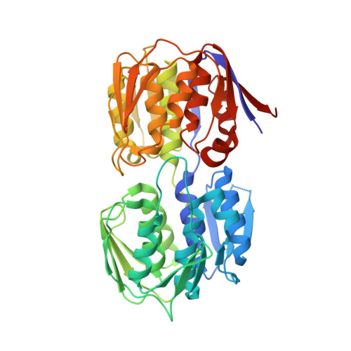
    MurA (enolpyruvyl UDP-GlcNAc synthase) catalyzes the first committed step in peptidoglycan biosynthesis. In this study, MurA-catalyzed breakdown of its tetrahedral intermediate (THI), with a k(cat)/K(M) of 520 M(-1) s(-1), was far slower than the normal reaction, and 3 x 10(5)-fold slower than the homologous enzyme, AroA, reacting with its THI. This provided kinetic evidence of slow binding and a conformationally constrained active site. The MurA cocrystal structure with UDP-N-acetylmuramic acid (UDP-MurNAc), a potent inhibitor, and phosphite revealed a new "staged" MurA conformation in which the Arg397 side chain tracked phosphite out of the catalytic site. The closed-to-staged transition involved breaking eight MurA.ligand ion pairs, and three intraprotein hydrogen bonds helping hold the active site loop closed. These were replaced with only two MurA.UDP-MurNAc ion pairs, two with phosphite, and seven new intraprotein ion pairs or hydrogen bonds. Cys115 appears to have an important role in forming the staged conformation. The staged conformation appears to be one step in a complex choreography of release of the product from MurA.


  • Organizational Affiliation
    • Department of Biochemistry and Biomedical Sciences, McMaster University, 1280 Main Street West, Hamilton,Ontario L8S 4M1, Canada.

Macromolecule Content 

  • Total Structure Weight: 546 kDa 
  • Atom Count: 39,025 
  • Modeled Residue Count: 5,016 
  • Deposited Residue Count: 5,016 
  • Unique protein chains: 1

Macromolecules

Find similar proteins by:|  3D Structure
Entity ID: 1
MoleculeChains  Sequence LengthOrganismDetailsImage
UDP-N-acetylglucosamine 1-carboxyvinyltransferase
A, B, C, D, E
A, B, C, D, E, F, G, H, I, J, K, L
418Escherichia coli K-12Mutation(s): 1 
Gene Names: b3189JW3156murAmurZ
EC: 2.5.1.7
UniProt
Find proteins for P0A749 (Escherichia coli (strain K12))
Explore P0A749 
Go to UniProtKB:  P0A749
Entity Groups
Sequence Clusters30% Identity50% Identity70% Identity90% Identity95% Identity100% Identity
UniProt GroupP0A749
Sequence Annotations
Expand
Reference Sequence

Small Molecules

Ligands 2 Unique
IDChains Name / Formula / InChI Key2D Diagram3D Interactions
EPU

Query on EPU


Download:Ideal Coordinates CCD File
BA [auth H]
DA [auth I]
FA [auth J]
HA [auth K]
JA [auth L]
BA [auth H],
DA [auth I],
FA [auth J],
HA [auth K],
JA [auth L],
N [auth A],
P [auth B],
R [auth C],
T [auth D],
V [auth E],
X [auth F],
Z [auth G]
URIDINE-DIPHOSPHATE-2(N-ACETYLGLUCOSAMINYL) BUTYRIC ACID
C20 H29 N3 O19 P2
BEGZZYPUNCJHKP-DBYWSUQTSA-N
PO3

Query on PO3


Download:Ideal Coordinates CCD File
AA [auth H]
CA [auth I]
EA [auth J]
GA [auth K]
IA [auth L]
AA [auth H],
CA [auth I],
EA [auth J],
GA [auth K],
IA [auth L],
M [auth A],
O [auth B],
Q [auth C],
S [auth D],
U [auth E],
W [auth F],
Y [auth G]
PHOSPHITE ION
O3 P
AQSJGOWTSHOLKH-UHFFFAOYSA-N

Experimental Data & Validation

Experimental Data

  • Method: X-RAY DIFFRACTION
  • Resolution: 2.50 Å
  • R-Value Free:  0.254 (Depositor), 0.249 (DCC) 
  • R-Value Work:  0.222 (Depositor), 0.217 (DCC) 
  • R-Value Observed: 0.223 (Depositor) 
Space Group: P 1
Unit Cell:
Length ( Å )Angle ( ˚ )
a = 84.51α = 111.52
b = 120.91β = 104.44
c = 139.73γ = 90.19
Software Package:
Software NamePurpose
ADSCdata collection
PHASERphasing
PHENIXrefinement
d*TREKdata reduction
d*TREKdata scaling

Structure Validation

View Full Validation Report


Ligand Structure Quality Assessment 


Entry History 

Deposition Data

Revision History  (Full details and data files)

  • Version 1.0: 2009-11-24
    Type: Initial release
  • Version 1.1: 2011-07-13
    Changes: Version format compliance
  • Version 1.2: 2011-07-27
    Changes: Database references, Derived calculations, Non-polymer description
  • Version 1.3: 2021-10-13
    Changes: Database references, Derived calculations
  • Version 1.4: 2023-09-06
    Changes: Data collection, Refinement description