3IQI | pdb_00003iqi

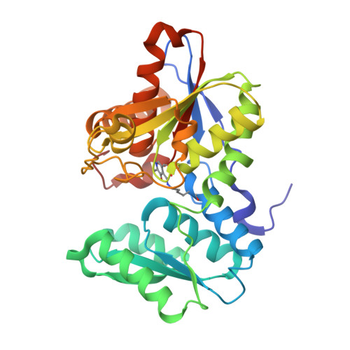
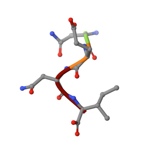
Structure of O-Acetylserine Sulfhydrylase in Complex with Peptide MNENI


Experimental Data Snapshot

  • Method: X-RAY DIFFRACTION
  • Resolution: 1.70 Å
  • R-Value Free: 
    0.204 (Depositor), 0.202 (DCC) 
  • R-Value Work: 
    0.172 (Depositor), 0.169 (DCC) 
  • R-Value Observed: 
    0.174 (Depositor) 

Starting Model: experimental
View more details

wwPDB Validation 3D Report Full Report

Validation slider image for 3IQI

This is version 1.3 of the entry. See complete history. 

Literature

Design of o-acetylserine sulfhydrylase inhibitors by mimicking nature.

Salsi, E., Bayden, A.S., Spyrakis, F., Amadasi, A., Campanini, B., Bettati, S., Dodatko, T., Cozzini, P., Kellogg, G.E., Cook, P.F., Roderick, S.L., Mozzarelli, A.

(2010) J Med Chem 53: 345-356

  • DOI: https://doi.org/10.1021/jm901325e
  • Primary Citation Related Structures: 
    3IQG, 3IQH, 3IQI

  • PubMed Abstract: 

    The inhibition of cysteine biosynthesis in prokaryotes and protozoa has been proposed to be relevant for the development of antibiotics. Haemophilus influenzae O-acetylserine sulfhydrylase (OASS), catalyzing l-cysteine formation, is inhibited by the insertion of the C-terminal pentapeptide (MNLNI) of serine acetyltransferase into the active site. Four-hundred MNXXI pentapeptides were generated in silico, docked into OASS active site using GOLD, and scored with HINT. The terminal P5 Ile accounts for about 50% of the binding energy. Glu or Asp at position P4 and, to a lesser extent, at position P3 also significantly contribute to the binding interaction. The predicted affinity of 14 selected pentapeptides correlated well with the experimentally determined dissociation constants. The X-ray structure of three high affinity pentapeptide-OASS complexes were compared with the docked poses. These results, combined with a GRID analysis of the active site, allowed us to define a pharmacophoric scaffold for the design of peptidomimetic inhibitors.


  • Organizational Affiliation: 
    • Department of Biochemistry and Molecular Biology, University of Parma, Italy. andrea.mozzarelli@unipr.it

Macromolecule Content 

  • Total Structure Weight: 34.27 kDa 
  • Atom Count: 2,597 
  • Modeled Residue Count: 314 
  • Deposited Residue Count: 321 
  • Unique protein chains: 2

Macromolecules

Find similar proteins by:|  3D Structure
Entity ID: 1
MoleculeChains  Sequence LengthOrganismDetailsImage
Cysteine synthaseA [auth X]316Haemophilus influenzaeMutation(s): 0 
Gene Names: cysK, HI1103
EC: 2.5.1.47
UniProt
Find proteins for P45040 (Haemophilus influenzae (strain ATCC 51907 / DSM 11121 / KW20 / Rd))
Explore P45040 
Go to UniProtKB:  P45040
Entity Groups
Sequence Clusters30% Identity50% Identity70% Identity90% Identity95% Identity100% Identity
UniProt GroupP45040
Sequence Annotations
Expand
Reference Sequence
Find similar proteins by:  Sequence   |   3D Structure  
Entity ID: 2
MoleculeChains  Sequence LengthOrganismDetailsImage
MNENIB [auth P]5N/AMutation(s): 0 
Sequence Annotations
Expand
Reference Sequence

Small Molecules

Modified Residues  1 Unique
IDChains TypeFormula2D DiagramParent
LLP
Query on LLP
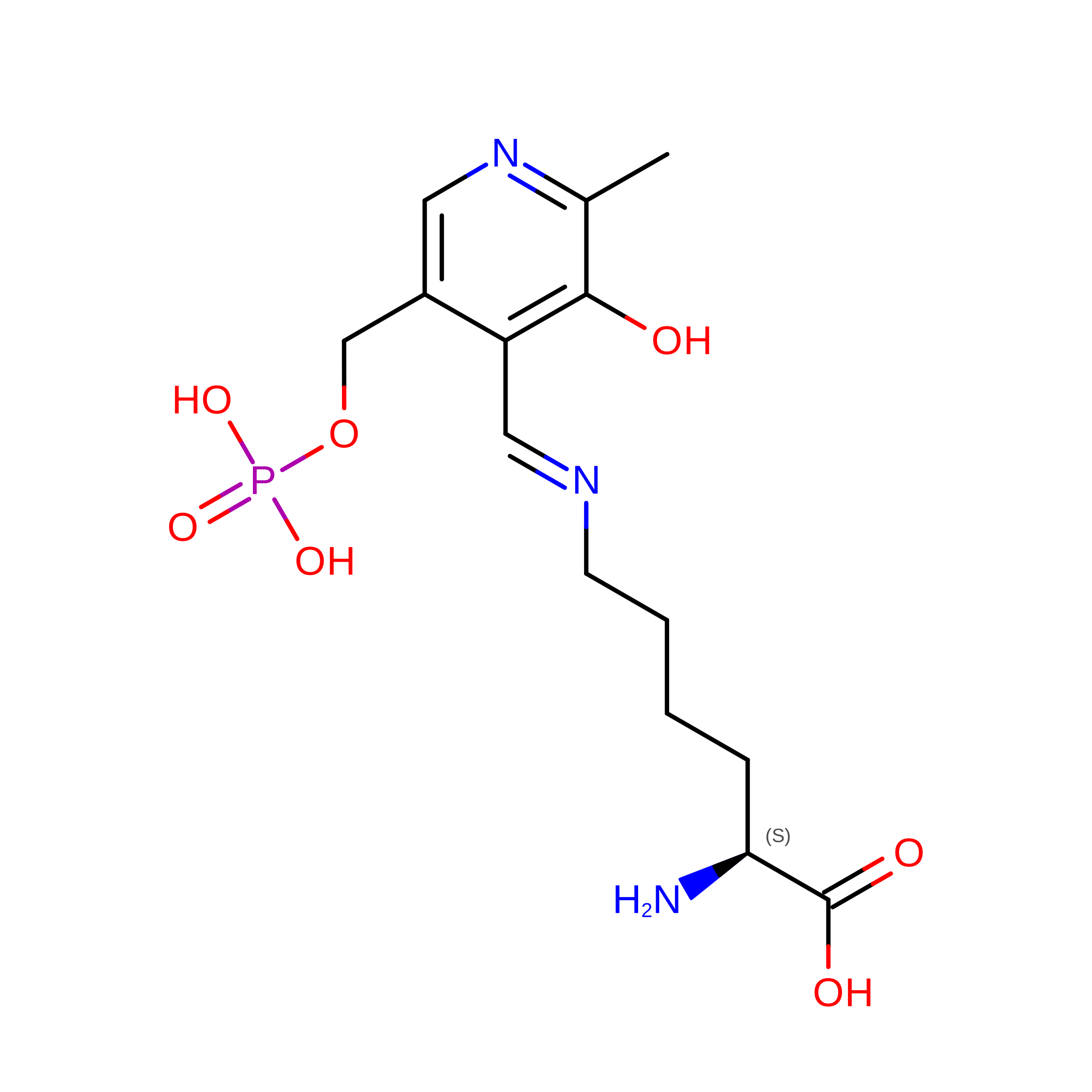
A [auth X]L-PEPTIDE LINKINGC14 H22 N3 O7 PLYS

Experimental Data & Validation

Experimental Data

  • Method: X-RAY DIFFRACTION
  • Resolution: 1.70 Å
  • R-Value Free:  0.204 (Depositor), 0.202 (DCC) 
  • R-Value Work:  0.172 (Depositor), 0.169 (DCC) 
  • R-Value Observed: 0.174 (Depositor) 
Space Group: I 41
Unit Cell:
Length ( Å )Angle ( ˚ )
a = 112.264α = 90
b = 112.264β = 90
c = 45.835γ = 90
Software Package:
Software NamePurpose
StructureStudiodata collection
REFMACrefinement
MOSFLMdata reduction
SCALAdata scaling
REFMACphasing

Structure Validation

View Full Validation Report


Entry History 

Deposition Data

Revision History  (Full details and data files)

  • Version 1.0: 2009-11-17
    Type: Initial release
  • Version 1.1: 2011-07-13
    Changes: Version format compliance
  • Version 1.2: 2023-09-06
    Changes: Data collection, Database references, Derived calculations, Refinement description
  • Version 1.3: 2023-11-22
    Changes: Data collection