3IOF | pdb_00003iof

Crystal structure of CphA N220G mutant with inhibitor 10a


Experimental Data Snapshot

  • Method: X-RAY DIFFRACTION
  • Resolution: 1.44 Å
  • R-Value Free: 
    0.141 (Depositor), 0.164 (DCC) 
  • R-Value Work: 
    0.123 (Depositor), 0.146 (DCC) 
  • R-Value Observed: 
    0.124 (Depositor) 

Starting Model: experimental
View more details

wwPDB Validation 3D Report Full Report

Validation slider image for 3IOF

Ligand Structure Quality Assessment 


This is version 1.3 of the entry. See complete history

Literature

Mercaptophosphonate Compounds as Broad-Spectrum Inhibitors of the Metallo-beta-lactamases

Lassaux, P.Hamel, M.Gulea, M.Delbruck, H.Mercuri, P.S.Horsfall, L.Dehareng, D.Kupper, M.Frere, J.-M.Hoffmann, K.Galleni, M.Bebrone, C.

(2010) J Med Chem 53: 4862-4876

  • DOI: https://doi.org/10.1021/jm100213c
  • Primary Citation Related Structures: 
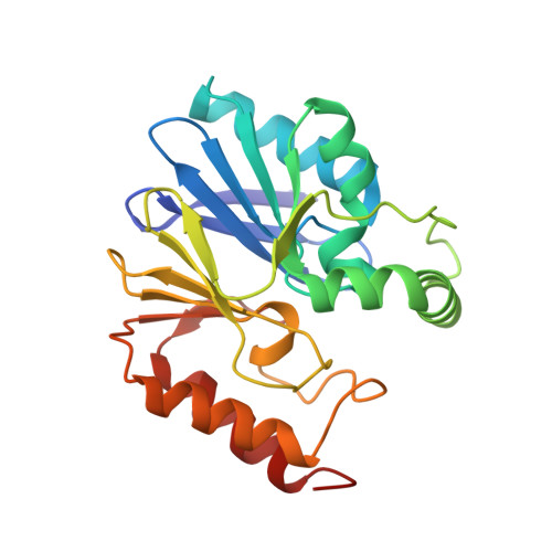
    2WRS, 3IOF, 3IOG

  • PubMed Abstract: 

    Although commercialized inhibitors of active site serine beta-lactamases are currently used in coadministration with antibiotic therapy, no clinically useful inhibitors of metallo-beta-lactamases (MBLs) have yet been discovered. In this paper, we investigated the inhibitory effect of mercaptophosphonate derivatives against the three subclasses of MBLs (B1, B2, and B3). All 14 tested mercaptophosphonates, with the exception of 1a, behaved as competitive inhibitors for the three subclasses. Apart from 13 and 21, all the mercaptophosphonates tested exhibit a good inhibitory effect on the subclass B2 MBL CphA with low inhibition constants (K(i) < 15 muM). Interestingly, compound 18 turned out to be a potent broad spectrum MBL inhibitor. The crystallographic structures of the CphA-10a and CphA-18 complexes indicated that the sulfur atom of 10a and the phosphonato group of 18 interact with the Zn(2+) ion, respectively. Molecular modeling studies of the interactions between compounds 10a and 18 and the VIM-4 (B1), CphA (B2), and FEZ-1 (B3) enzymes brought to light different binding modes depending on the enzyme and the inhibitor, consistent with the crystallographic structures.


  • Organizational Affiliation
    • Laboratory of Biological Macromolecules, University of Liège, Allée du 6 Août B6, Sart-Tilman, 4000 Liège, Belgium.

Macromolecule Content 

  • Total Structure Weight: 26.75 kDa 
  • Atom Count: 2,309 
  • Modeled Residue Count: 227 
  • Deposited Residue Count: 227 
  • Unique protein chains: 1

Macromolecules

Find similar proteins by:|  3D Structure
Entity ID: 1
MoleculeChains  Sequence LengthOrganismDetailsImage
Beta-lactamase227Aeromonas hydrophilaMutation(s): 1 
Gene Names: cphA
EC: 3.5.2.6
UniProt
Find proteins for P26918 (Aeromonas hydrophila)
Explore P26918 
Go to UniProtKB:  P26918
Entity Groups
Sequence Clusters30% Identity50% Identity70% Identity90% Identity95% Identity100% Identity
UniProt GroupP26918
Sequence Annotations
Expand
Reference Sequence

Small Molecules

Ligands 4 Unique
IDChains Name / Formula / InChI Key2D Diagram3D Interactions
IFS

Query on IFS


Download:Ideal Coordinates CCD File
C [auth A]bis(1-methylethyl) [2-(sulfanylmethyl)phenyl]phosphonate
C13 H21 O3 P S
JFZVPWMZPZJUTE-UHFFFAOYSA-N
SO4

Query on SO4


Download:Ideal Coordinates CCD File
D [auth A]
E [auth A]
F [auth A]
G [auth A]
H [auth A]
D [auth A],
E [auth A],
F [auth A],
G [auth A],
H [auth A],
I [auth A],
J [auth A],
K [auth A],
L [auth A],
M [auth A]
SULFATE ION
O4 S
QAOWNCQODCNURD-UHFFFAOYSA-L
GOL

Query on GOL


Download:Ideal Coordinates CCD File
N [auth A],
O [auth A],
P [auth A]
GLYCEROL
C3 H8 O3
PEDCQBHIVMGVHV-UHFFFAOYSA-N
ZN

Query on ZN


Download:Ideal Coordinates CCD File
B [auth A]ZINC ION
Zn
PTFCDOFLOPIGGS-UHFFFAOYSA-N
Binding Affinity Annotations 
IDSourceBinding Affinity
IFS BindingDB:  3IOF Ki: 2000 (nM) from 1 assay(s)

Experimental Data & Validation

Experimental Data

  • Method: X-RAY DIFFRACTION
  • Resolution: 1.44 Å
  • R-Value Free:  0.141 (Depositor), 0.164 (DCC) 
  • R-Value Work:  0.123 (Depositor), 0.146 (DCC) 
  • R-Value Observed: 0.124 (Depositor) 
Space Group: C 2 2 21
Unit Cell:
Length ( Å )Angle ( ˚ )
a = 42.65α = 90
b = 100.66β = 90
c = 116.99γ = 90
Software Package:
Software NamePurpose
MAR345dtbdata collection
PHASERphasing
REFMACrefinement
XDSdata reduction
SCALAdata scaling

Structure Validation

View Full Validation Report


Ligand Structure Quality Assessment 


Entry History 

Deposition Data

Revision History  (Full details and data files)

  • Version 1.0: 2010-06-23
    Type: Initial release
  • Version 1.1: 2011-07-13
    Changes: Version format compliance
  • Version 1.2: 2015-01-14
    Changes: Refinement description
  • Version 1.3: 2023-11-01
    Changes: Data collection, Database references, Derived calculations, Refinement description