


Crystal structure of pantoate-beta-alanine-ligase in complex with ATP at low occupancy at 2.1 A resolution
Seattle Structural Genomics Center for Infectious Disease (SSGCID), Abendroth, J., Davies, D., Staker, B.To be published.
Experimental Data Snapshot
Starting Model: experimental
View more details
Entity ID: 1 | |||||
|---|---|---|---|---|---|
| Molecule | Chains | Sequence Length | Organism | Details | Image |
| Pantothenate synthetase | 314 | Brucella melitensis | Mutation(s): 0 Gene Names: BMEI1593, panC EC: 6.3.2.1 | 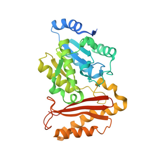 | |
UniProt | |||||
Entity Groups | |||||
| Sequence Clusters | 30% Identity50% Identity70% Identity90% Identity95% Identity100% Identity | ||||
| UniProt Group | Q8YFC9 | ||||
Sequence AnnotationsExpand | |||||
Reference Sequence | |||||
| Ligands 2 Unique | |||||
|---|---|---|---|---|---|
| ID | Chains | Name / Formula / InChI Key | 2D Diagram | 3D Interactions | |
| ATP Download:Ideal Coordinates CCD File | E [auth A], I [auth C], M [auth D] | ADENOSINE-5'-TRIPHOSPHATE C10 H16 N5 O13 P3 ZKHQWZAMYRWXGA-KQYNXXCUSA-N |  | ||
| UNX Download:Ideal Coordinates CCD File | F [auth A] G [auth A] H [auth A] J [auth C] K [auth C] | UNKNOWN ATOM OR ION X |  | ||
| Length ( Å ) | Angle ( ˚ ) |
|---|---|
| a = 94.32 | α = 90 |
| b = 94.32 | β = 90 |
| c = 236.93 | γ = 120 |
| Software Name | Purpose |
|---|---|
| Blu-Ice | data collection |
| PHASER | phasing |
| REFMAC | refinement |
| XDS | data reduction |
| XSCALE | data scaling |


