3IKN

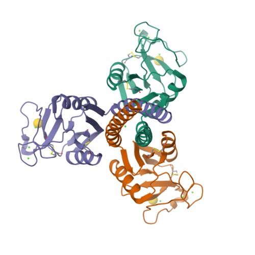
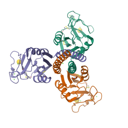
Crystal structure of galactose bound trimeric human lung surfactant protein D


Experimental Data Snapshot

  • Method: X-RAY DIFFRACTION
  • Resolution: 1.60 Å
  • R-Value Free: 0.215 
  • R-Value Work: 0.201 

Starting Model: experimental
View more details

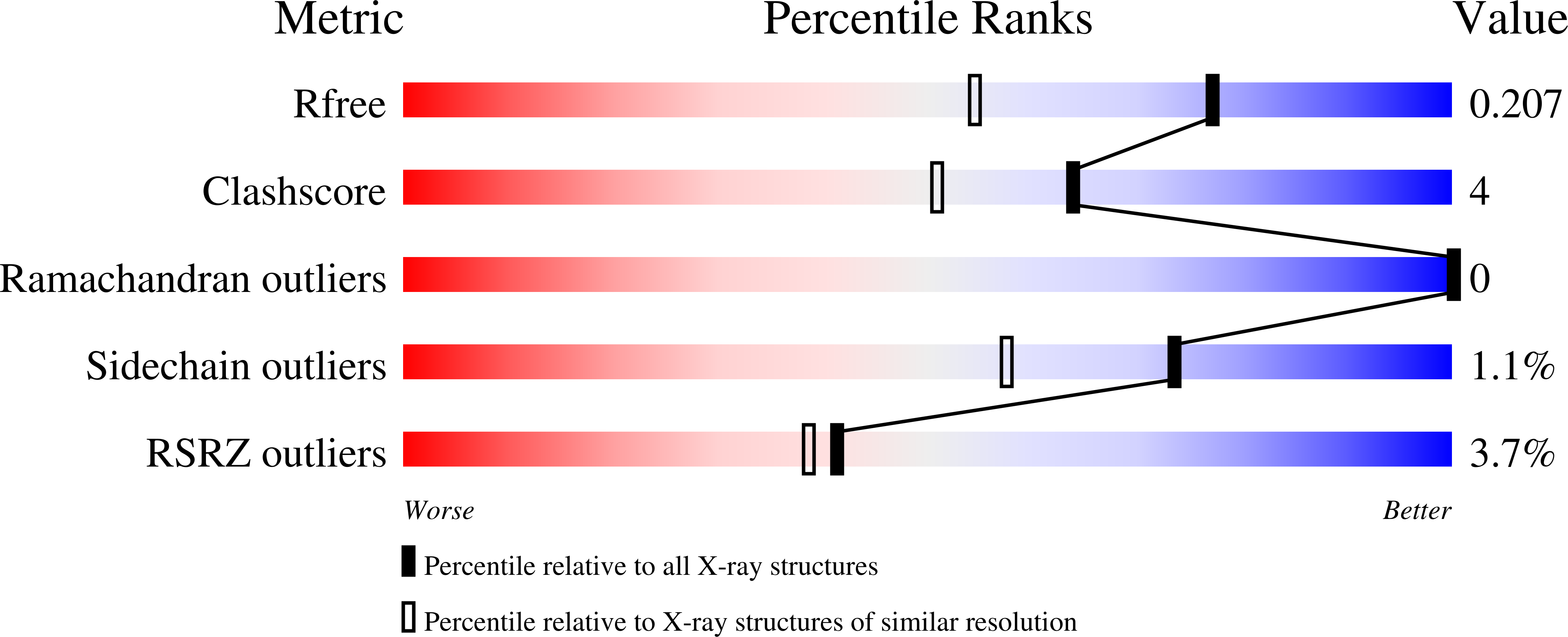
wwPDB Validation   3D Report Full Report


Ligand Structure Quality Assessment 


This is version 1.6 of the entry. See complete history


Literature

Structural characterisation of ligand-binding determinants in human lung surfactant protein D: influence of Asp325

Shrive, A.K.Martin, C.Burns, I.Paterson, J.M.Martin, J.D.Townsend, J.P.Waters, P.Clark, H.W.Kishore, U.Reid, K.B.M.Greenhough, T.J.

(2009) J Mol Biol 394: 776-788

  • DOI: https://doi.org/10.1016/j.jmb.2009.09.057
  • Primary Citation of Related Structures:  
    3IKN, 3IKP, 3IKQ, 3IKR

  • PubMed Abstract: 

    The crystal structures of a biologically and therapeutically active recombinant homotrimeric fragment of human lung surfactant protein D with a series of bound ligands have been determined. While the structures reveal various different binding modes, all utilise a similarly positioned pair of mannose-type O3' and O4' hydroxyls with no direct interaction between any non-terminal sugar and protein. The orientation, position, and interactions of the bound terminal sugar depend on the sugar itself, the presence and form of glycosidic linkage, and the environment in the crystal, which, via Asp325, places stereochemical and electronic constraints, different for the three different subunits in the homotrimer, on the ligand-binding site. As a direct consequence of this influence, the other binding-pocket flanking residue, Arg343, exhibits variable conformation and variable interactions with bound ligand and leaves open to question which orientation of terminal mannobiose, and of other terminal disaccharides, may be present in extended physiological ligands. The combined structural evidence shows that there is significant flexibility in recognition; that Asp325, in addition to Arg343, is an important determinant of ligand selectivity, recognition, and binding; and that differences in crystal contact interfaces exert, through Asp325, significant influence on preferred binding modes.


  • Organizational Affiliation

    Research Institute of Science and Technology in Medicine, and School of Life Sciences, Keele University, Staffordshire ST5 5BG, UK. a.k.shrive@keele.ac.uk


Macromolecules
Find similar proteins by:  (by identity cutoff)  |  3D Structure
Entity ID: 1
MoleculeChains Sequence LengthOrganismDetailsImage
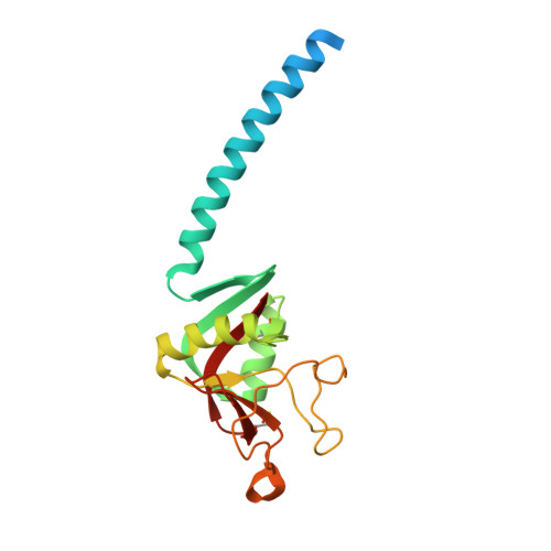
Pulmonary surfactant-associated protein D
A, B, C
177Homo sapiensMutation(s): 1 
Gene Names: SFTPDPSPDSFTP4
UniProt & NIH Common Fund Data Resources
Find proteins for P35247 (Homo sapiens)
Explore P35247 
Go to UniProtKB:  P35247
PHAROS:  P35247
GTEx:  ENSG00000133661 
Entity Groups  
Sequence Clusters30% Identity50% Identity70% Identity90% Identity95% Identity100% Identity
UniProt GroupP35247
Sequence Annotations
Expand
  • Reference Sequence
Small Molecules
Ligands 2 Unique
IDChains Name / Formula / InChI Key2D Diagram3D Interactions
GAL
Query on GAL

Download Ideal Coordinates CCD File 
H [auth A],
L [auth B],
P [auth C]
beta-D-galactopyranose
C6 H12 O6
WQZGKKKJIJFFOK-FPRJBGLDSA-N
CA
Query on CA

Download Ideal Coordinates CCD File 
D [auth A]
E [auth A]
F [auth A]
G [auth A]
I [auth B]
D [auth A],
E [auth A],
F [auth A],
G [auth A],
I [auth B],
J [auth B],
K [auth B],
M [auth C],
N [auth C],
O [auth C]
CALCIUM ION
Ca
BHPQYMZQTOCNFJ-UHFFFAOYSA-N
Experimental Data & Validation

Experimental Data

  • Method: X-RAY DIFFRACTION
  • Resolution: 1.60 Å
  • R-Value Free: 0.215 
  • R-Value Work: 0.201 
  • Space Group: P 1 21 1
Unit Cell:
Length ( Å )Angle ( ˚ )
a = 55.74α = 90
b = 108.58β = 91.41
c = 55.88γ = 90
Software Package:
Software NamePurpose
MOSFLMdata reduction
SCALAdata scaling
CNSrefinement
PDB_EXTRACTdata extraction
ADSCdata collection

Structure Validation

View Full Validation Report


Ligand Structure Quality Assessment 


Entry History 

Deposition Data

Revision History  (Full details and data files)

  • Version 1.0: 2009-11-17
    Type: Initial release
  • Version 1.1: 2011-07-13
    Changes: Version format compliance
  • Version 1.2: 2014-02-19
    Changes: Database references
  • Version 1.3: 2020-07-29
    Type: Remediation
    Reason: Carbohydrate remediation
    Changes: Data collection, Derived calculations, Structure summary
  • Version 1.4: 2021-11-10
    Changes: Database references, Structure summary
  • Version 1.5: 2023-11-01
    Changes: Data collection, Refinement description
  • Version 1.6: 2024-11-06
    Changes: Structure summary