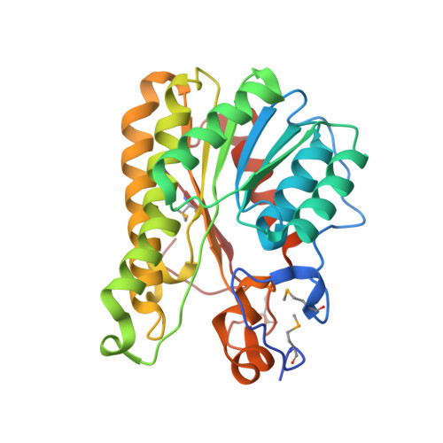
2.05 Angstrom resolution crystal structure of a short chain dehydrogenase from Bacillus anthracis str. 'Ames Ancestor' in complex with NAD+
Halavaty, A.S., Minasov, G., Skarina, T., Onopriyenko, O., Gordon, E., Kwon, K., Savchenko, A., Anderson, W.F., Center for Structural Genomics of Infectious Diseases (CSGID)To be published.