3IJF | pdb_00003ijf

Crystal structure of cytidine deaminase from Mycobacterium tuberculosis


Experimental Data Snapshot

  • Method: X-RAY DIFFRACTION
  • Resolution: 1.99 Å
  • R-Value Free: 
    0.240 (Depositor), 0.224 (DCC) 
  • R-Value Work: 
    0.190 (Depositor) 
  • R-Value Observed: 
    0.193 (Depositor) 

Starting Model: experimental
View more details

wwPDB Validation 3D Report Full Report

Validation slider image for 3IJF

This is version 1.2 of the entry. See complete history

Literature

Structural and functional analyses of Mycobacterium tuberculosis Rv3315c-encoded metal-dependent homotetrameric cytidine deaminase.

Sanchez-Quitian, Z.A.Schneider, C.Z.Ducati, R.G.de Azevedo, W.F.Bloch, C.Basso, L.A.Santos, D.S.

(2010) J Struct Biol 169: 413-423

  • DOI: https://doi.org/10.1016/j.jsb.2009.12.019
  • Primary Citation Related Structures: 
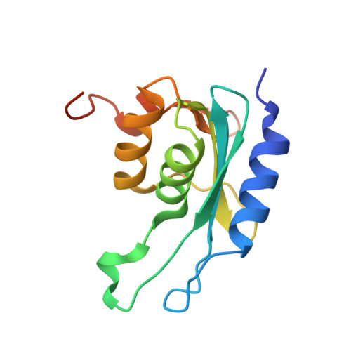
    3IJF

  • PubMed Abstract: 

    The emergence of drug-resistant strains of Mycobacterium tuberculosis, the causative agent of tuberculosis, has exacerbated the treatment and control of this disease. Cytidine deaminase (CDA) is a pyrimidine salvage pathway enzyme that recycles cytidine and 2'-deoxycytidine for uridine and 2'-deoxyuridine synthesis, respectively. A probable M. tuberculosis CDA-coding sequence (cdd, Rv3315c) was cloned, sequenced, expressed in Escherichia coli BL21(DE3), and purified to homogeneity. Mass spectrometry, N-terminal amino acid sequencing, gel filtration chromatography, and metal analysis of M. tuberculosis CDA (MtCDA) were carried out. These results and multiple sequence alignment demonstrate that MtCDA is a homotetrameric Zn(2+)-dependent metalloenzyme. Steady-state kinetic measurements yielded the following parameters: K(m)=1004 microM and k(cat)=4.8s(-1) for cytidine, and K(m)=1059 microM and k(cat)=3.5s(-1) for 2'-deoxycytidine. The pH dependence of k(cat) and k(cat)/K(M) for cytidine indicate that protonation of a single ionizable group with apparent pK(a) value of 4.3 abolishes activity, and protonation of a group with pK(a) value of 4.7 reduces binding. MtCDA was crystallized and crystal diffracted at 2.0 A resolution. Analysis of the crystallographic structure indicated the presence of a Zn(2+) coordinated by three conserved cysteines and the structure exhibits the canonical cytidine deaminase fold.


  • Organizational Affiliation
    • Centro de Pesquisas em Biologia Molecular e Funcional (CPBMF), Instituto Nacional de Ciência e Tecnologia em Tuberculose (INCT-TB), Pontifícia Universidade Católica do Rio Grande do Sul (PUCRS), Av. Ipiranga, 6681, Porto Alegre, RS 90619-900, Brazil.

Macromolecule Content 

  • Total Structure Weight: 14.15 kDa 
  • Atom Count: 983 
  • Modeled Residue Count: 123 
  • Deposited Residue Count: 133 
  • Unique protein chains: 1

Macromolecules

Find similar proteins by:|  3D Structure
Entity ID: 1
MoleculeChains  Sequence LengthOrganismDetailsImage
Cytidine deaminaseA [auth X]133Mycobacterium tuberculosisMutation(s): 0 
Gene Names: cddMT3416Rv3315c
EC: 3.5.4.5
UniProt
Find proteins for P9WPH3 (Mycobacterium tuberculosis (strain ATCC 25618 / H37Rv))
Explore P9WPH3 
Go to UniProtKB:  P9WPH3
Entity Groups
Sequence Clusters30% Identity50% Identity70% Identity90% Identity95% Identity100% Identity
UniProt GroupP9WPH3
Sequence Annotations
Expand
Reference Sequence

Small Molecules

Ligands 1 Unique
IDChains Name / Formula / InChI Key2D Diagram3D Interactions
ZN

Query on ZN


Download:Ideal Coordinates CCD File
B [auth X]ZINC ION
Zn
PTFCDOFLOPIGGS-UHFFFAOYSA-N

Experimental Data & Validation

Experimental Data

  • Method: X-RAY DIFFRACTION
  • Resolution: 1.99 Å
  • R-Value Free:  0.240 (Depositor), 0.224 (DCC) 
  • R-Value Work:  0.190 (Depositor) 
  • R-Value Observed: 0.193 (Depositor) 
Space Group: C 2 2 2
Unit Cell:
Length ( Å )Angle ( ˚ )
a = 63.705α = 90
b = 75.338β = 90
c = 55.116γ = 90
Software Package:
Software NamePurpose
HKL-3000data collection
AMoREphasing
REFMACrefinement
MOSFLMdata reduction
SCALAdata scaling

Structure Validation

View Full Validation Report


Entry History 

Deposition Data

Revision History  (Full details and data files)

  • Version 1.0: 2010-03-02
    Type: Initial release
  • Version 1.1: 2011-07-13
    Changes: Version format compliance
  • Version 1.2: 2023-09-06
    Changes: Data collection, Database references, Derived calculations, Refinement description