3II9 | pdb_00003ii9

Crystal structure of glutaryl-coa dehydrogenase from Burkholderia pseudomallei at 1.73 Angstrom


Experimental Data Snapshot

  • Method: X-RAY DIFFRACTION
  • Resolution: 1.74 Å
  • R-Value Free: 
    0.196 (Depositor), 0.248 (DCC) 
  • R-Value Work: 
    0.170 (Depositor) 
  • R-Value Observed: 
    0.172 (Depositor) 

Starting Model: experimental
View more details

wwPDB Validation 3D Report Full Report

Validation slider image for 3II9

This is version 1.3 of the entry. See complete history

Literature

User-loaded SlipChip for equipment-free multiplexed nanoliter-scale experiments.

Li, L.Du, W.Ismagilov, R.

(2010) J Am Chem Soc 132: 106-111

  • DOI: https://doi.org/10.1021/ja908555n
  • Primary Citation Related Structures: 
    3II9

  • PubMed Abstract: 

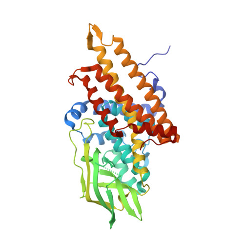
    This paper describes a microfluidic approach to perform multiplexed nanoliter-scale experiments by combining a sample with multiple different reagents, each at multiple mixing ratios. This approach employs a user-loaded, equipment-free SlipChip. The mixing ratios, characterized by diluting a fluorescent dye, could be controlled by the volume of each of the combined wells. The SlipChip design was validated on an approximately 12 nL scale by screening the conditions for crystallization of glutaryl-CoA dehydrogenase from Burkholderia pseudomallei against 48 different reagents; each reagent was tested at 11 different mixing ratios, for a total of 528 crystallization trials. The total consumption of the protein sample was approximately 10 microL. Conditions for crystallization were successfully identified. The crystallization experiments were successfully scaled up in well plates using the conditions identified in the SlipChip. Crystals were characterized by X-ray diffraction and provided a protein structure in a different space group and at a higher resolution than the structure obtained by conventional methods. In this work, this user-loaded SlipChip has been shown to reliably handle fluids of diverse physicochemical properties, such as viscosities and surface tensions. Quantitative measurements of fluorescent intensities and high-resolution imaging were straighforward to perform in these glass SlipChips. Surface chemistry was controlled using fluorinated lubricating fluid, analogous to the fluorinated carrier fluid used in plug-based crystallization. Thus, we expect this approach to be valuable in a number of areas beyond protein crystallization, especially those areas where droplet-based microfluidic systems have demonstrated successes, including measurements of enzyme kinetics and blood coagulation, cell-based assays, and chemical reactions.


  • Organizational Affiliation
    • Department of Chemistry and Institute for Biophysical Dynamics, The University of Chicago, 929 East 57th Street, Chicago, Illinois 60637, USA.

Macromolecule Content 

  • Total Structure Weight: 174.89 kDa 
  • Atom Count: 12,893 
  • Modeled Residue Count: 1,540 
  • Deposited Residue Count: 1,584 
  • Unique protein chains: 1

Macromolecules

Find similar proteins by:|  3D Structure
Entity ID: 1
MoleculeChains  Sequence LengthOrganismDetailsImage
Glutaryl-CoA dehydrogenase
A, B, C, D
396Burkholderia pseudomallei 1710bMutation(s): 0 
Gene Names: BURPS1710b_3237
EC: 1.3.99.7 (PDB Primary Data), 1.3.8.6 (UniProt)
UniProt
Find proteins for Q3JP94 (Burkholderia pseudomallei (strain 1710b))
Explore Q3JP94 
Go to UniProtKB:  Q3JP94
Entity Groups
Sequence Clusters30% Identity50% Identity70% Identity90% Identity95% Identity100% Identity
UniProt GroupQ3JP94
Sequence Annotations
Expand
Reference Sequence

Small Molecules

Ligands 5 Unique
IDChains Name / Formula / InChI Key2D Diagram3D Interactions
PG4

Query on PG4


Download:Ideal Coordinates CCD File
H [auth B],
I [auth B],
P [auth C]
TETRAETHYLENE GLYCOL
C8 H18 O5
UWHCKJMYHZGTIT-UHFFFAOYSA-N
PGE

Query on PGE


Download:Ideal Coordinates CCD File
G [auth A],
K [auth C]
TRIETHYLENE GLYCOL
C6 H14 O4
ZIBGPFATKBEMQZ-UHFFFAOYSA-N
PEG

Query on PEG


Download:Ideal Coordinates CCD File
E [auth A],
L [auth C],
S [auth D],
U [auth D]
DI(HYDROXYETHYL)ETHER
C4 H10 O3
MTHSVFCYNBDYFN-UHFFFAOYSA-N
GOL

Query on GOL


Download:Ideal Coordinates CCD File
J [auth B],
N [auth C],
Q [auth C],
R [auth D],
T [auth D]
GLYCEROL
C3 H8 O3
PEDCQBHIVMGVHV-UHFFFAOYSA-N
MG

Query on MG


Download:Ideal Coordinates CCD File
F [auth A],
M [auth C],
O [auth C],
V [auth D]
MAGNESIUM ION
Mg
JLVVSXFLKOJNIY-UHFFFAOYSA-N

Experimental Data & Validation

Experimental Data

  • Method: X-RAY DIFFRACTION
  • Resolution: 1.74 Å
  • R-Value Free:  0.196 (Depositor), 0.248 (DCC) 
  • R-Value Work:  0.170 (Depositor) 
  • R-Value Observed: 0.172 (Depositor) 
Space Group: P 1 21 1
Unit Cell:
Length ( Å )Angle ( ˚ )
a = 80.966α = 90
b = 141.268β = 112.26
c = 84.012γ = 90
Software Package:
Software NamePurpose
HKL-2000data collection
AMoREphasing
REFMACrefinement
HKL-2000data reduction
HKL-2000data scaling

Structure Validation

View Full Validation Report


Entry History 

Revision History  (Full details and data files)

  • Version 1.0: 2009-12-15
    Type: Initial release
  • Version 1.1: 2011-07-13
    Changes: Advisory, Version format compliance
  • Version 1.2: 2012-10-10
    Changes: Database references
  • Version 1.3: 2023-09-06
    Changes: Data collection, Database references, Derived calculations, Refinement description