3IGI | pdb_00003igi

Tertiary Architecture of the Oceanobacillus Iheyensis Group II Intron


Experimental Data Snapshot

  • Method: X-RAY DIFFRACTION
  • Resolution: 3.13 Å
  • R-Value Free: 
    0.218 (Depositor), 0.208 (DCC) 
  • R-Value Work: 
    0.183 (Depositor), 0.176 (DCC) 
  • R-Value Observed: 
    0.184 (Depositor) 

Starting Model: experimental
View more details

wwPDB Validation 3D Report Full Report

Validation slider image for 3IGI

This is version 1.3 of the entry. See complete history

Literature

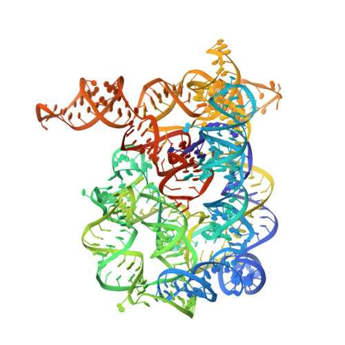
Tertiary architecture of the Oceanobacillus iheyensis group II intron.

Toor, N.Keating, K.S.Fedorova, O.Rajashankar, K.Wang, J.Pyle, A.M.

(2010) RNA 16: 57-69

  • DOI: https://doi.org/10.1261/rna.1844010
  • Primary Citation Related Structures: 
    3IGI

  • PubMed Abstract: 

    Group II introns are large ribozymes that act as self-splicing and retrotransposable RNA molecules. They are of great interest because of their potential evolutionary relationship to the eukaryotic spliceosome, their continued influence on the organization of many genomes in bacteria and eukaryotes, and their potential utility as tools for gene therapy and biotechnology. One of the most interesting features of group II introns is their relative lack of nucleobase conservation and covariation, which has long suggested that group II intron structures are stabilized by numerous unusual tertiary interactions and backbone-mediated contacts. Here, we provide a detailed description of the tertiary interaction networks within the Oceanobacillus iheyensis group IIC intron, for which a crystal structure was recently solved to 3.1 A resolution. The structure can be described as a set of several intricately constructed tertiary interaction nodes, each of which contains a core of extended stacking networks and elaborate motifs. Many of these nodes are surrounded by a web of ribose zippers, which appear to further stabilize local structure. As predicted from biochemical and genetic studies, the group II intron provides a wealth of new information on strategies for RNA folding and tertiary structural organization.


  • Organizational Affiliation
    • Department of Molecular Biophysics and Biochemistry, Yale University, New Haven, Connecticut 06511, USA.

Macromolecule Content 

  • Total Structure Weight: 139.6 kDa 
  • Atom Count: 8,656 
  • Modeled Residue Count: 398 
  • Deposited Residue Count: 425 
  • Unique nucleic acid chains: 2

Macromolecules

Find similar nucleic acids by:  (by identity cutoff) 
Entity ID: 1
MoleculeChains LengthOrganismImage
Group IIC intron412N/A
Sequence Annotations
Expand
Reference Sequence
Find similar nucleic acids by:  Sequence
Entity ID: 2
MoleculeChains LengthOrganismImage
5'-R(*CP*GP*CP*UP*CP*UP*AP*CP*UP*CP*UP*AP*U)-3'13N/A
Sequence Annotations
Expand
Reference Sequence

Small Molecules

Ligands 2 Unique
IDChains Name / Formula / InChI Key2D Diagram3D Interactions
K

Query on K


Download:Ideal Coordinates CCD File
AB [auth A]
BB [auth A]
CB [auth A]
DB [auth A]
EB [auth A]
AB [auth A],
BB [auth A],
CB [auth A],
DB [auth A],
EB [auth A],
FB [auth A],
GB [auth A],
HB [auth A],
IB [auth A],
JB [auth A],
KB [auth A],
LB [auth A],
MB [auth A],
NB [auth A],
OB [auth A],
PB [auth A],
UA [auth A],
VA [auth A],
WA [auth A],
XA [auth A],
YA [auth A],
ZA [auth A]
POTASSIUM ION
K
NPYPAHLBTDXSSS-UHFFFAOYSA-N
MG

Query on MG


Download:Ideal Coordinates CCD File
AA [auth A]
BA [auth A]
C [auth A]
CA [auth A]
D [auth A]
AA [auth A],
BA [auth A],
C [auth A],
CA [auth A],
D [auth A],
DA [auth A],
E [auth A],
EA [auth A],
F [auth A],
FA [auth A],
G [auth A],
GA [auth A],
H [auth A],
HA [auth A],
I [auth A],
IA [auth A],
J [auth A],
JA [auth A],
K [auth A],
KA [auth A],
L [auth A],
LA [auth A],
M [auth A],
MA [auth A],
N [auth A],
NA [auth A],
O [auth A],
OA [auth A],
P [auth A],
PA [auth A],
Q [auth A],
QA [auth A],
R [auth A],
RA [auth A],
S [auth A],
SA [auth A],
T [auth A],
TA [auth A],
U [auth A],
V [auth A],
W [auth A],
X [auth A],
Y [auth A],
Z [auth A]
MAGNESIUM ION
Mg
JLVVSXFLKOJNIY-UHFFFAOYSA-N

Experimental Data & Validation

Experimental Data

  • Method: X-RAY DIFFRACTION
  • Resolution: 3.13 Å
  • R-Value Free:  0.218 (Depositor), 0.208 (DCC) 
  • R-Value Work:  0.183 (Depositor), 0.176 (DCC) 
  • R-Value Observed: 0.184 (Depositor) 
Space Group: P 21 21 21
Unit Cell:
Length ( Å )Angle ( ˚ )
a = 89.092α = 90
b = 94.969β = 90
c = 225.972γ = 90
Software Package:
Software NamePurpose
HKL-2000data collection
SHELXmodel building
PHENIXrefinement
HKL-2000data reduction
HKL-2000data scaling
SHELXphasing

Structure Validation

View Full Validation Report


Entry History 

Deposition Data

Revision History  (Full details and data files)

  • Version 1.0: 2009-12-22
    Type: Initial release
  • Version 1.1: 2011-07-13
    Changes: Version format compliance
  • Version 1.2: 2013-09-25
    Changes: Derived calculations
  • Version 1.3: 2023-09-06
    Changes: Data collection, Database references, Derived calculations, Refinement description