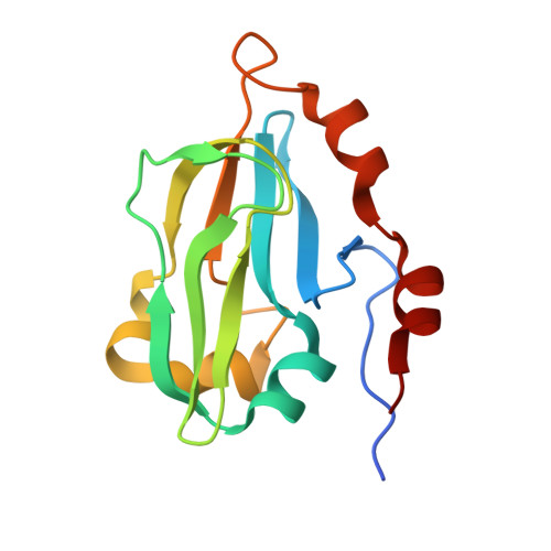
3IFT | pdb_00003ift

Crystal structure of glycine cleavage system protein H from Mycobacterium tuberculosis, using X-rays from the Compact Light Source.


Experimental Data Snapshot

  • Method: X-RAY DIFFRACTION
  • Resolution: 2.00 Å
  • R-Value Free: 
    0.253 (Depositor), 0.251 (DCC) 
  • R-Value Work: 
    0.181 (Depositor), 0.183 (DCC) 
  • R-Value Observed: 
    0.185 (Depositor) 

Starting Model: experimental
View more details

wwPDB Validation 3D Report Full Report

Validation slider image for 3IFT

This is version 1.3 of the entry. See complete history. 

Literature

X-ray structure determination of the glycine cleavage system protein H of Mycobacterium tuberculosis using an inverse Compton synchrotron X-ray source.

Abendroth, J., McCormick, M.S., Edwards, T.E., Staker, B., Loewen, R., Gifford, M., Rifkin, J., Mayer, C., Guo, W., Zhang, Y., Myler, P., Kelley, A., Analau, E., Hewitt, S.N., Napuli, A.J., Kuhn, P., Ruth, R.D., Stewart, L.J.

(2010) J Struct Funct Genomics 11: 91-100

  • DOI: https://doi.org/10.1007/s10969-010-9087-6
  • Primary Citation Related Structures: 
    3HGB, 3IFT

  • PubMed Abstract: 

    Structural genomics discovery projects require ready access to both X-ray diffraction and NMR spectroscopy which support the collection of experimental data needed to solve large numbers of novel protein structures. The most productive X-ray crystal structure determination laboratories make extensive use of tunable synchrotron X-ray light to solve novel structures by anomalous diffraction methods. This requires that frozen cryo-protected crystals be shipped to large multi acre synchrotron facilities for data collection. In this paper we report on the development and use of the first laboratory-scale synchrotron light source capable of performing many of the state-of-the-art synchrotron applications in X-ray science. This Compact Light Source is a first-in-class device that uses inverse Compton scattering to generate X-rays of sufficient flux, tunable wavelength and beam size to allow high-resolution X-ray diffraction data collection from protein crystals. We report on benchmarking tests of X-ray diffraction data collection with hen egg white lysozyme, and the successful high-resolution X-ray structure determination of the Glycine cleavage system protein H from Mycobacterium tuberculosis using diffraction data collected with the Compact Light Source X-ray beam.


  • Organizational Affiliation: 
    • Emerald BioStructures, 7869 NE Day Road West, Bainbridge Island, WA 98110, USA.

Macromolecule Content 

  • Total Structure Weight: 15.24 kDa 
  • Atom Count: 1,165 
  • Modeled Residue Count: 136 
  • Deposited Residue Count: 142 
  • Unique protein chains: 1

Macromolecules

Find similar proteins by:|  3D Structure
Entity ID: 1
MoleculeChains  Sequence LengthOrganismDetailsImage
Glycine cleavage system H protein142Mycobacterium tuberculosisMutation(s): 0 
Gene Names: GCHV, gcvH, MT1874, MTCY1A11.17c, Rv1826
UniProt
Find proteins for P9WN55 (Mycobacterium tuberculosis (strain ATCC 25618 / H37Rv))
Explore P9WN55 
Go to UniProtKB:  P9WN55
Entity Groups
Sequence Clusters30% Identity50% Identity70% Identity90% Identity95% Identity100% Identity
UniProt GroupP9WN55
Sequence Annotations
Expand
Reference Sequence

Experimental Data & Validation

Experimental Data

  • Method: X-RAY DIFFRACTION
  • Resolution: 2.00 Å
  • R-Value Free:  0.253 (Depositor), 0.251 (DCC) 
  • R-Value Work:  0.181 (Depositor), 0.183 (DCC) 
  • R-Value Observed: 0.185 (Depositor) 
Space Group: C 1 2 1
Unit Cell:
Length ( Å )Angle ( ˚ )
a = 86.45α = 90
b = 51.01β = 95.1
c = 32.57γ = 90
Software Package:
Software NamePurpose
MOLREPphasing
REFMACrefinement
XDSdata reduction
XSCALEdata scaling

Structure Validation

View Full Validation Report


Entry History 

Revision History  (Full details and data files)

  • Version 1.0: 2009-08-11
    Type: Initial release
  • Version 1.1: 2011-07-13
    Changes: Version format compliance
  • Version 1.2: 2018-04-04
    Changes: Data collection
  • Version 1.3: 2023-09-06
    Changes: Data collection, Database references, Refinement description