Pyrazole-based cathepsin S inhibitors with arylalkynes as P1 binding elements.
Ameriks, M.K., Axe, F.U., Bembenek, S.D., Edwards, J.P., Gu, Y., Karlsson, L., Randal, M., Sun, S., Thurmond, R.L., Zhu, J.(2009) Bioorg Med Chem Lett 19: 6131-6134
- PubMed: 19773165 Search on PubMed
- DOI: https://doi.org/10.1016/j.bmcl.2009.09.014
- Primary Citation Related Structures:
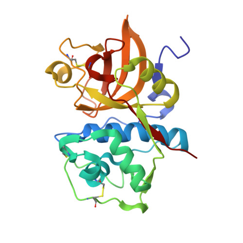
3IEJ - PubMed Abstract:
A crystal structure of 1 bound to a Cys25Ser mutant of cathepsin S helped to elucidate the binding mode of a previously disclosed series of pyrazole-based CatS inhibitors and facilitated the design of a new class of arylalkyne analogs. Optimization of the alkyne and tetrahydropyridine portions of the pharmacophore provided potent CatS inhibitors (IC50=40-300 nM), and an X-ray structure of 32 revealed that the arylalkyne moiety binds in the S1 pocket of the enzyme.
- Johnson & Johnson Pharmaceutical Research & Development, L.L.C., 3210 Merryfield Row, San Diego, CA 92121, USA. mameriks@its.jnj.com
Organizational Affiliation: