3IBH | pdb_00003ibh

Crystal structure of Saccharomyces cerevisiae Gtt2 in complex with glutathione


Experimental Data Snapshot

  • Method: X-RAY DIFFRACTION
  • Resolution: 2.10 Å
  • R-Value Free: 
    0.247 (Depositor), 0.246 (DCC) 
  • R-Value Work: 
    0.208 (Depositor), 0.207 (DCC) 
  • R-Value Observed: 
    0.210 (Depositor) 

Starting Model: experimental
View more details

wwPDB Validation 3D Report Full Report

Validation slider image for 3IBH

Ligand Structure Quality Assessment 


This is version 1.3 of the entry. See complete history

Literature

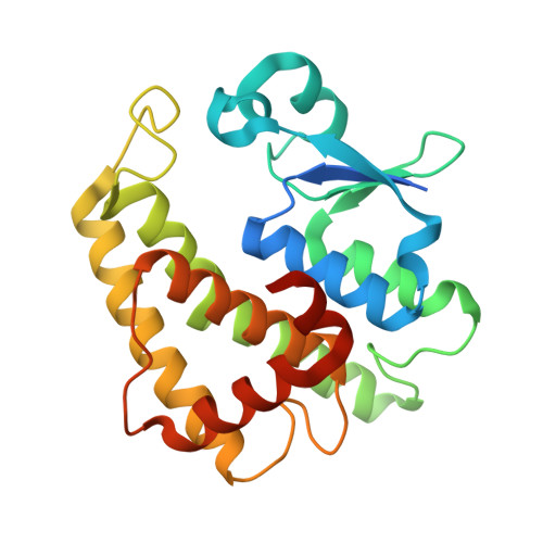
Structures of yeast glutathione-S-transferase Gtt2 reveal a new catalytic type of GST family.

Ma, X.X.Jiang, Y.L.He, Y.X.Bao, R.Chen, Y.X.Zhou, C.Z.

(2009) EMBO Rep 10: 1320-1326

  • DOI: https://doi.org/10.1038/embor.2009.216
  • Primary Citation Related Structures: 
    3ERF, 3ERG, 3IBH

  • PubMed Abstract: 

    Glutathione-S-transferases (GSTs) are ubiquitous detoxification enzymes that catalyse the conjugation of electrophilic substrates to glutathione. Here, we present the crystal structures of Gtt2, a GST of Saccharomyces cerevisiae, in apo and two ligand-bound forms, at 2.23 A, 2.20 A and 2.10 A, respectively. Although Gtt2 has the overall structure of a GST, the absence of the classic catalytic essential residues--tyrosine, serine and cysteine--distinguishes it from all other cytosolic GSTs of known structure. Site-directed mutagenesis in combination with activity assays showed that instead of the classic catalytic residues, a water molecule stabilized by Ser129 and His123 acts as the deprotonator of the glutathione sulphur atom. Furthermore, only glycine and alanine are allowed at the amino-terminus of helix-alpha1 because of stereo-hindrance. Taken together, these results show that yeast Gtt2 is a novel atypical type of cytosolic GST.


  • Organizational Affiliation
    • Hefei National Laboratory for Physical Sciences at Microscale, and School of Life Sciences, University of Science and Technology of China, Hefei, Anhui 230027, People's Republic of China.

Macromolecule Content 

  • Total Structure Weight: 26.68 kDa 
  • Atom Count: 1,789 
  • Modeled Residue Count: 208 
  • Deposited Residue Count: 233 
  • Unique protein chains: 1

Macromolecules

Find similar proteins by:|  3D Structure
Entity ID: 1
MoleculeChains  Sequence LengthOrganismDetailsImage
Saccharomyces cerevisiae Gtt2233Saccharomyces cerevisiaeMutation(s): 0 
Gene Names: GTT2L0560YLL060C
EC: 2.5.1.18
UniProt
Find proteins for Q12390 (Saccharomyces cerevisiae (strain ATCC 204508 / S288c))
Explore Q12390 
Go to UniProtKB:  Q12390
Entity Groups
Sequence Clusters30% Identity50% Identity70% Identity90% Identity95% Identity100% Identity
UniProt GroupQ12390
Sequence Annotations
Expand
Reference Sequence

Small Molecules

Ligands 1 Unique
IDChains Name / Formula / InChI Key2D Diagram3D Interactions
GSH

Query on GSH


Download:Ideal Coordinates CCD File
B [auth A]Glutathione
C10 H17 N3 O6 S
RWSXRVCMGQZWBV-WDSKDSINSA-N

Biologically Interesting Molecules (External Reference) 

1 Unique

Experimental Data & Validation

Experimental Data

  • Method: X-RAY DIFFRACTION
  • Resolution: 2.10 Å
  • R-Value Free:  0.247 (Depositor), 0.246 (DCC) 
  • R-Value Work:  0.208 (Depositor), 0.207 (DCC) 
  • R-Value Observed: 0.210 (Depositor) 
Space Group: P 31 2 1
Unit Cell:
Length ( Å )Angle ( ˚ )
a = 88.774α = 90
b = 88.774β = 90
c = 69.102γ = 120
Software Package:
Software NamePurpose
MAR345dtbdata collection
AMoREphasing
REFMACrefinement
HKL-2000data reduction
HKL-2000data scaling

Structure Validation

View Full Validation Report


Ligand Structure Quality Assessment 


Entry History 

Deposition Data

Revision History  (Full details and data files)

  • Version 1.0: 2009-10-13
    Type: Initial release
  • Version 1.1: 2011-07-13
    Changes: Version format compliance
  • Version 1.2: 2011-12-07
    Changes: Non-polymer description
  • Version 1.3: 2023-11-01
    Changes: Data collection, Database references, Derived calculations, Refinement description