3I8D | pdb_00003i8d

The Pairing Geometry of the Hydrophobic Thymine Analog 2,4-Difluorotoluene in Duplex DNA as Analyzed by X-ray Crystallography


Experimental Data Snapshot

  • Method: X-RAY DIFFRACTION
  • Resolution: 1.61 Å
  • R-Value Free: 
    0.238 (Depositor), 0.237 (DCC) 
  • R-Value Work: 
    0.197 (Depositor), 0.195 (DCC) 
  • R-Value Observed: 
    0.199 (Depositor) 

Starting Model: experimental
View more details

wwPDB Validation 3D Report Full Report

Validation slider image for 3I8D

This is version 1.4 of the entry. See complete history

Literature

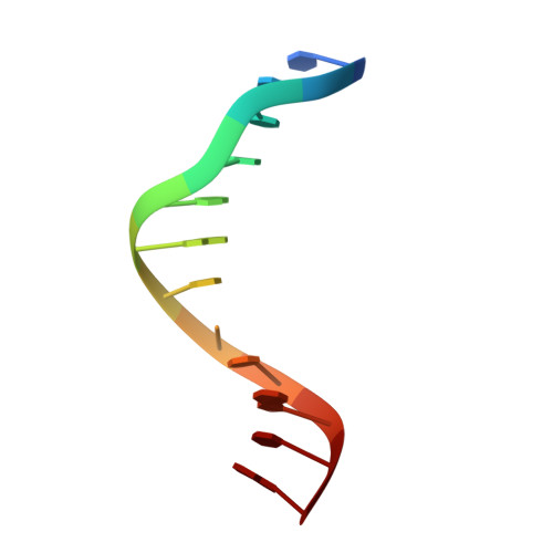
Pairing geometry of the hydrophobic thymine analogue 2,4-difluorotoluene in duplex DNA as analyzed by X-ray crystallography.

Pallan, P.S.Egli, M.

(2009) J Am Chem Soc 131: 12548-12549

  • DOI: https://doi.org/10.1021/ja905739j
  • Primary Citation Related Structures: 
    3I8D

  • PubMed Abstract: 

    Certain DNA polymerases (pols) were found to efficiently insert A opposite the hydrophobic T isostere 2,4-difluorotoluene (F) and vice versa, resulting in the widely held belief that some pols rely on shape rather than H-bonding for accurate replication. Using X-ray crystallography we have analyzed the geometry of F:A pairs in duplex DNA and observed a distance between fluorine and the exocyclic amino group of A that is consistent with a H-bond, thus challenging the assumption that the F analogue is unable to engage in H-bonding as well as the steric hypothesis of DNA replication. Therefore, shape and H-bonding are inherently related, and steric constraints at a pol active site, or conferred by stacking or the DNA backbone conformation, may enable H-bonding by F.


  • Organizational Affiliation
    • Department of Biochemistry, School of Medicine, Vanderbilt University, Nashville, Tennessee 37232, USA.

Macromolecule Content 

  • Total Structure Weight: 38.19 kDa 
  • Atom Count: 2,895 
  • Modeled Residue Count: 288 
  • Deposited Residue Count: 288 
  • Unique protein chains: 1
  • Unique nucleic acid chains: 1

Macromolecules


Find similar proteins by:|  3D Structure
Entity ID: 1
MoleculeChains  Sequence LengthOrganismDetailsImage
Ribonuclease H
A, C
132Halalkalibacterium haloduransMutation(s): 1 
Gene Names: BACILLUS HALODURANSBH0863RNase-H D132N mutantrnhA
EC: 3.1.26.4
UniProt
Find proteins for Q9KEI9 (Halalkalibacterium halodurans (strain ATCC BAA-125 / DSM 18197 / FERM 7344 / JCM 9153 / C-125))
Explore Q9KEI9 
Go to UniProtKB:  Q9KEI9
Entity Groups
Sequence Clusters30% Identity50% Identity70% Identity90% Identity95% Identity100% Identity
UniProt GroupQ9KEI9
Sequence Annotations
Expand
Reference Sequence
Find similar nucleic acids by:  Sequence
Entity ID: 2
MoleculeChains LengthOrganismImage
5'-D(*CP*GP*CP*GP*AP*AP*TP*(DFT)P*CP*GP*CP*G)-3'
B, D
12N/A
Sequence Annotations
Expand
Reference Sequence

Experimental Data & Validation

Experimental Data

  • Method: X-RAY DIFFRACTION
  • Resolution: 1.61 Å
  • R-Value Free:  0.238 (Depositor), 0.237 (DCC) 
  • R-Value Work:  0.197 (Depositor), 0.195 (DCC) 
  • R-Value Observed: 0.199 (Depositor) 
Space Group: C 1 2 1
Unit Cell:
Length ( Å )Angle ( ˚ )
a = 96.178α = 90
b = 66.714β = 121.04
c = 77.571γ = 90
Software Package:
Software NamePurpose
MAR345data collection
MOLREPphasing
REFMACrefinement
HKL-2000data reduction
HKL-2000data scaling

Structure Validation

View Full Validation Report


Entry History 

Deposition Data

Revision History  (Full details and data files)

  • Version 1.0: 2009-09-01
    Type: Initial release
  • Version 1.1: 2011-07-13
    Changes: Advisory, Refinement description, Version format compliance
  • Version 1.2: 2017-11-01
    Changes: Advisory, Refinement description
  • Version 1.3: 2021-10-13
    Changes: Advisory, Database references, Derived calculations
  • Version 1.4: 2023-09-06
    Changes: Data collection, Refinement description