3I6N | pdb_00003i6n

Mode of Binding of the Tuberculosis Prodrug Isoniazid to Peroxidases: Crystal Structure of Bovine Lactoperoxidase with Isoniazid at 2.7 Resolution


Experimental Data Snapshot

  • Method: X-RAY DIFFRACTION
  • Resolution: 2.70 Å
  • R-Value Free: 
    0.232 (Depositor), 0.220 (DCC) 
  • R-Value Work: 
    0.205 (Depositor) 
  • R-Value Observed: 
    0.205 (Depositor) 

Starting Model: experimental
View more details

wwPDB Validation 3D Report Full Report

Validation slider image for 3I6N

Ligand Structure Quality Assessment 


This is version 2.2 of the entry. See complete history

Literature

Mode of binding of the tuberculosis prodrug isoniazid to heme peroxidases: binding studies and crystal structure of bovine lactoperoxidase with isoniazid at 2.7 A resolution.

Singh, A.K.Kumar, R.P.Pandey, N.Singh, N.Sinha, M.Bhushan, A.Kaur, P.Sharma, S.Singh, T.P.

(2010) J Biological Chem 285: 1569-1576

  • DOI: https://doi.org/10.1074/jbc.M109.060327
  • Primary Citation Related Structures: 
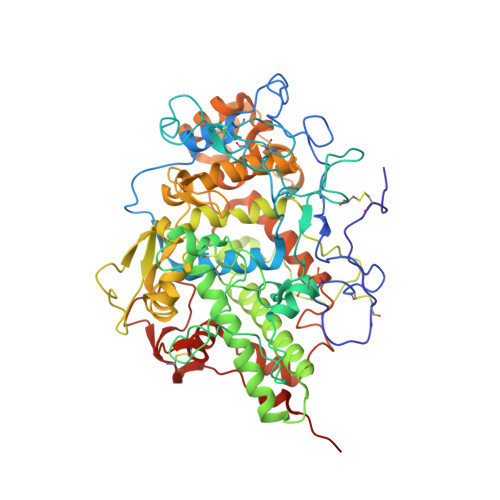
    3GC1, 3I6N

  • PubMed Abstract: 

    Isoniazid (INH) is an anti-tuberculosis prodrug that is activated by mammalian lactoperoxidase and Mycobacterium tuberculosis catalase peroxidase (MtCP). We report here binding studies, an enzyme assay involving INH, and the crystal structure of the complex of bovine lactoperoxidase (LPO) with INH to illuminate binding properties and INH activation as well as the mode of diffusion and interactions together with a detailed structural and functional comparison with MtCP. The structure determination shows that isoniazid binds to LPO at the substrate binding site on the distal heme side. The substrate binding site is connected to the protein surface through a long hydrophobic channel. The acyl hydrazide moiety of isoniazid interacts with Phe(422) O, Gln(423) O(epsilon1), and Phe(254) O. In this arrangement, pyridinyl nitrogen forms a hydrogen bond with a water molecule, W-1, which in turn forms three hydrogen bonds with Fe(3+), His(109) N(epsilon2), and Gln(105) N(epsilon2). The remaining two sides of isoniazid form hydrophobic interactions with the atoms of heme pyrrole ring A, C(beta) and C(gamma) atoms of Glu(258), and C(gamma) and C(delta) atoms of Arg(255). The binding studies indicate that INH binds to LPO with a value of 0.9 x 10(-6) m for the dissociation constant. The nitro blue tetrazolium reduction assay shows that INH is activated by the reaction of LPO-H(2)O(2) with INH. This suggests that LPO can be used for INH activation. It also indicates that the conversion of INH into isonicotinoyl radical by LPO may be the cause of INH toxicity.


  • Organizational Affiliation
    • Department of Biophysics, All India Institute of Medical Sciences, Ansari Nagar, New Delhi 110 029, India.

Macromolecule Content 

  • Total Structure Weight: 71.61 kDa 
  • Atom Count: 5,259 
  • Modeled Residue Count: 595 
  • Deposited Residue Count: 595 
  • Unique protein chains: 1

Macromolecules

Find similar proteins by:|  3D Structure
Entity ID: 1
MoleculeChains  Sequence LengthOrganismDetailsImage
Lactoperoxidase595Bos taurusMutation(s): 0 
EC: 1.11.1.7
UniProt
Find proteins for P80025 (Bos taurus)
Explore P80025 
Go to UniProtKB:  P80025
Entity Groups
Sequence Clusters30% Identity50% Identity70% Identity90% Identity95% Identity100% Identity
UniProt GroupP80025
Glycosylation
Glycosylation Sites: 4
Sequence Annotations
Expand
Reference Sequence

Oligosaccharides

Help  
Entity ID: 2
MoleculeChains Length2D Diagram GlycosylationD Interactions
alpha-D-mannopyranose-(1-4)-2-acetamido-2-deoxy-beta-D-glucopyranose-(1-4)-2-acetamido-2-deoxy-beta-D-glucopyranose
B, D
3N-Glycosylation
Glycosylation Resources
GlyTouCan: G62182OO
GlyCosmos: G62182OO
GlyGen: G62182OO
Entity ID: 3
MoleculeChains Length2D Diagram GlycosylationD Interactions
2-acetamido-2-deoxy-beta-D-glucopyranose-(1-4)-2-acetamido-2-deoxy-beta-D-glucopyranose
C, E
2N-Glycosylation
Glycosylation Resources
GlyTouCan: G42666HT
GlyCosmos: G42666HT
GlyGen: G42666HT

Small Molecules

Ligands 5 Unique
IDChains Name / Formula / InChI Key2D Diagram3D Interactions
HEM

Query on HEM


Download:Ideal Coordinates CCD File
F [auth A]PROTOPORPHYRIN IX CONTAINING FE
C34 H32 Fe N4 O4
KABFMIBPWCXCRK-RGGAHWMASA-L
ISZ

Query on ISZ


Download:Ideal Coordinates CCD File
G [auth A]4-(DIAZENYLCARBONYL)PYRIDINE
C6 H5 N3 O
QFLYISKABRUWCS-VQHVLOKHSA-N
IOD

Query on IOD


Download:Ideal Coordinates CCD File
H [auth A]
I [auth A]
J [auth A]
K [auth A]
L [auth A]
H [auth A],
I [auth A],
J [auth A],
K [auth A],
L [auth A],
M [auth A],
N [auth A]
IODIDE ION
I
XMBWDFGMSWQBCA-UHFFFAOYSA-M
SCN

Query on SCN


Download:Ideal Coordinates CCD File
P [auth A]THIOCYANATE ION
C N S
ZMZDMBWJUHKJPS-UHFFFAOYSA-M
CA

Query on CA


Download:Ideal Coordinates CCD File
O [auth A]CALCIUM ION
Ca
BHPQYMZQTOCNFJ-UHFFFAOYSA-N
Modified Residues  1 Unique
IDChains TypeFormula2D DiagramParent
SEP
Query on SEP
A
L-PEPTIDE LINKINGC3 H8 N O6 PSER

Experimental Data & Validation

Experimental Data

  • Method: X-RAY DIFFRACTION
  • Resolution: 2.70 Å
  • R-Value Free:  0.232 (Depositor), 0.220 (DCC) 
  • R-Value Work:  0.205 (Depositor) 
  • R-Value Observed: 0.205 (Depositor) 
Space Group: P 1 21 1
Unit Cell:
Length ( Å )Angle ( ˚ )
a = 54.49α = 90
b = 80.55β = 102.66
c = 77.78γ = 90
Software Package:
Software NamePurpose
DENZOdata reduction
AMoREphasing
CNSrefinement
HKL-2000data reduction
SCALEPACKdata scaling

Structure Validation

View Full Validation Report


Ligand Structure Quality Assessment 


Entry History 

Deposition Data

Revision History  (Full details and data files)

  • Version 1.0: 2009-10-13
    Type: Initial release
  • Version 1.1: 2011-07-13
    Changes: Non-polymer description, Version format compliance
  • Version 1.2: 2018-01-31
    Changes: Experimental preparation
  • Version 2.0: 2020-07-29
    Type: Remediation
    Reason: Carbohydrate remediation
    Changes: Atomic model, Data collection, Derived calculations, Structure summary
  • Version 2.1: 2023-11-01
    Changes: Data collection, Database references, Refinement description, Structure summary
  • Version 2.2: 2024-10-16
    Changes: Structure summary