


Crystal structure of muconate lactonizing enzyme from Ruegeria pomeroyi.
Fedorov, A.A., Fedorov, E.V., Sauder, J.M., Burley, S.K., Gerlt, J.A., Almo, S.C.To be published.
Experimental Data Snapshot
wwPDB Validation 3D Report Full Report

Entity ID: 1 | |||||
|---|---|---|---|---|---|
| Molecule | Chains | Sequence Length | Organism | Details | Image |
| Muconate cycloisomerase I | 385 | Ruegeria pomeroyi | Mutation(s): 0 Gene Names: catB, SPO3667 EC: 5.5.1.1 | 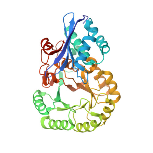 | |
UniProt | |||||
Entity Groups | |||||
| Sequence Clusters | 30% Identity50% Identity70% Identity90% Identity95% Identity100% Identity | ||||
| UniProt Group | Q5LM96 | ||||
Sequence AnnotationsExpand | |||||
Reference Sequence | |||||
| Ligands 2 Unique | |||||
|---|---|---|---|---|---|
| ID | Chains | Name / Formula / InChI Key | 2D Diagram | 3D Interactions | |
| MG Download:Ideal Coordinates CCD File | I [auth A] K [auth B] M [auth C] O [auth D] Q [auth E] | MAGNESIUM ION Mg JLVVSXFLKOJNIY-UHFFFAOYSA-N |  | ||
| NA Download:Ideal Coordinates CCD File | J [auth A] L [auth B] N [auth C] P [auth D] R [auth E] | SODIUM ION Na FKNQFGJONOIPTF-UHFFFAOYSA-N |  | ||
| Length ( Å ) | Angle ( ˚ ) |
|---|---|
| a = 109.135 | α = 90 |
| b = 135.358 | β = 103.18 |
| c = 113.024 | γ = 90 |
| Software Name | Purpose |
|---|---|
| ADSC | data collection |
| PHENIX | model building |
| CNS | refinement |
| DENZO | data reduction |
| SCALEPACK | data scaling |
| PHENIX | phasing |


