3I4W | pdb_00003i4w

Crystal Structure of the third PDZ domain of PSD-95


Experimental Data Snapshot

  • Method: X-RAY DIFFRACTION
  • Resolution: 1.35 Å
  • R-Value Free: 
    0.216 (Depositor), 0.216 (DCC) 
  • R-Value Work: 
    0.187 (Depositor), 0.188 (DCC) 
  • R-Value Observed: 
    0.189 (Depositor) 

Starting Model: experimental
View more details

wwPDB Validation 3D Report Full Report

Validation slider image for 3I4W

This is version 2.1 of the entry. See complete history

Literature

Novel conformational aspects of the third PDZ domain of the neuronal post-synaptic density-95 protein revealed from two 1.4A X-ray structures

Camara-Artigas, A.Murciano-Calles, J.Gavira, J.A.Cobos, E.S.Martinez, J.C.

(2010) J Struct Biol 170: 565-569

  • DOI: https://doi.org/10.1016/j.jsb.2010.03.005
  • Primary Citation Related Structures: 
    3I4W, 3K82

  • PubMed Abstract: 

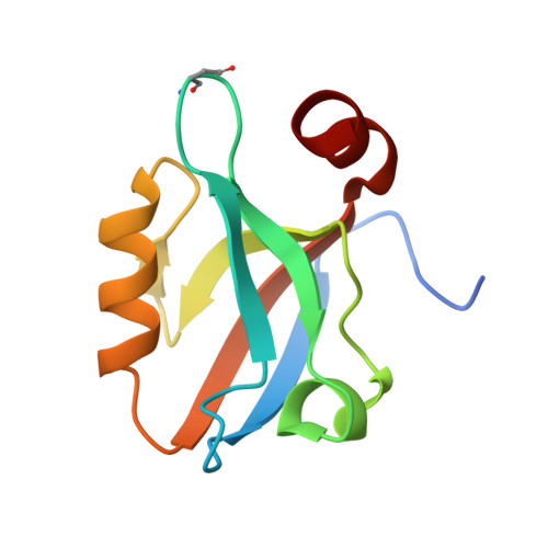
    The crystal structure of the third PDZ domain of the neuronal post-synaptic density-95 protein (PSD95-PDZ3, residues 302-402) has been solved at 1.4 and 1.35A from two different crystal forms. These structures lack the cloning artefact present in the carboxyl terminal sequence of the former crystallographic structures and they belong to the space groups P4(3) and P1. The new PDZ structures are identical between the two crystal forms and among the four chains of the P1 crystal form. When we compare the new structures with the previous ones, some important conformational differences in the C-terminal alpha-helix and in the loop connecting beta2 and beta3 strands have been found. Additionally, the high resolution of the new structures has allowed us to indentify a succinimide residue at the position corresponding to Asp332 in the beta2-beta3 loop, which may contribute to the alternate conformation of this loop, and at the same time, to the interaction between residues from this loop and the C-terminal alpha-helix. Thus, these features would have implications in the recently proposed allosteric role of this third alpha-helix in the binding of the carboxyl terminal fragments to the PSD95-PDZ3.


  • Organizational Affiliation
    • Department of Physical Chemistry, Biochemistry and Inorganic Chemistry, University of Almería, 04120 Almería, Spain. acamara@ual.es <acamara@ual.es>

Macromolecule Content 

  • Total Structure Weight: 44.87 kDa 
  • Atom Count: 3,241 
  • Modeled Residue Count: 392 
  • Deposited Residue Count: 416 
  • Unique protein chains: 1

Macromolecules

Find similar proteins by:|  3D Structure
Entity ID: 1
MoleculeChains  Sequence LengthOrganismDetailsImage
Disks large homolog 4
A, B, C, D
104Homo sapiensMutation(s): 1 
UniProt & NIH Common Fund Data Resources
Find proteins for P78352 (Homo sapiens)
Explore P78352 
Go to UniProtKB:  P78352
PHAROS:  P78352
GTEx:  ENSG00000132535 
Entity Groups
Sequence Clusters30% Identity50% Identity70% Identity90% Identity95% Identity100% Identity
UniProt GroupP78352
Sequence Annotations
Expand
Reference Sequence

Experimental Data & Validation

Experimental Data

  • Method: X-RAY DIFFRACTION
  • Resolution: 1.35 Å
  • R-Value Free:  0.216 (Depositor), 0.216 (DCC) 
  • R-Value Work:  0.187 (Depositor), 0.188 (DCC) 
  • R-Value Observed: 0.189 (Depositor) 
Space Group: P 1
Unit Cell:
Length ( Å )Angle ( ˚ )
a = 43.322α = 90
b = 42.152β = 90
c = 47.602γ = 92.08
Software Package:
Software NamePurpose
SCALAdata scaling
MOLREPphasing
REFMACrefinement
PDB_EXTRACTdata extraction
HKL-2000data collection
DENZOdata reduction
SCALEPACKdata scaling

Structure Validation

View Full Validation Report


Entry History 

Deposition Data

Revision History  (Full details and data files)

  • Version 1.0: 2010-04-07
    Type: Initial release
  • Version 1.1: 2011-07-13
    Changes: Version format compliance
  • Version 1.2: 2014-02-12
    Changes: Database references
  • Version 1.3: 2017-11-01
    Changes: Refinement description
  • Version 1.4: 2021-11-10
    Changes: Database references, Derived calculations
  • Version 1.5: 2023-11-01
    Changes: Data collection, Refinement description
  • Version 2.0: 2023-11-15
    Changes: Atomic model, Data collection, Derived calculations
  • Version 2.1: 2024-11-06
    Changes: Structure summary