3I32 | pdb_00003i32

Dimeric structure of a Hera helicase fragment including the C-terminal RecA domain, the dimerization domain, and the RNA binding domain


Experimental Data Snapshot

  • Method: X-RAY DIFFRACTION
  • Resolution: 2.80 Å
  • R-Value Free: 
    0.292 (Depositor), 0.306 (DCC) 
  • R-Value Work: 
    0.253 (Depositor), 0.265 (DCC) 
  • R-Value Observed: 
    0.255 (Depositor) 

Starting Model: experimental
View more details

wwPDB Validation 3D Report Full Report

Validation slider image for 3I32

This is version 1.4 of the entry. See complete history

Literature

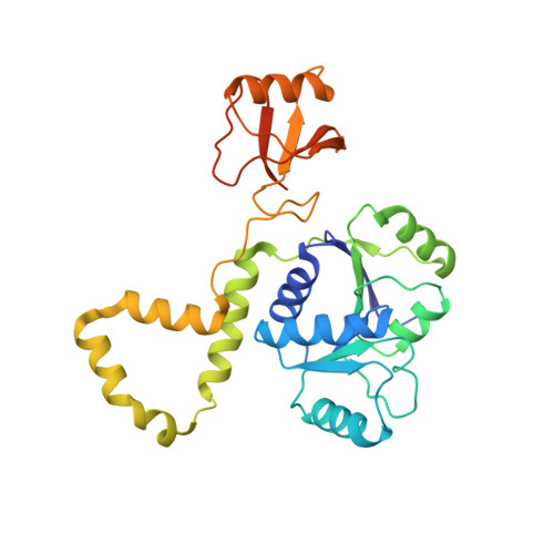
The Thermus thermophilus DEAD box helicase Hera contains a modified RNA recognition motif domain loosely connected to the helicase core.

Rudolph, M.G.Klostermeier, D.

(2009) RNA 15: 1993-2001

  • DOI: https://doi.org/10.1261/rna.1820009
  • Primary Citation Related Structures: 
    3I31, 3I32

  • PubMed Abstract: 

    DEAD box family helicases consist of a helicase core that is formed by two flexibly linked RecA-like domains. The helicase activity can be regulated by N- or C-terminal extensions flanking the core. Thermus thermophilus heat resistant RNA-dependent ATPase (Hera) is the first DEAD box helicase that forms a dimer using a unique dimerization domain. In addition to the dimerization domain, Hera contains a C-terminal RNA binding domain (RBD) that shares sequence homology only to uncharacterized proteins of the Deinococcus/Thermus group. The crystal structure of Hera_RBD reveals the fold of an altered RNA recognition motif (RRM) with limited structural homology to the RBD of the DEAD box helicase YxiN from Bacillus subtilis. Comparison with RRM/RNA complexes shows that a RNA binding mode different than that suggested for YxiN, but similar to U1A, can be inferred for Hera. The orientation of the RBD relative to the helicase core was defined in a second crystal structure of a Hera fragment including the C-terminal RecA domain, the dimerization domain, and the RBD. The structures allow construction of a model for the entire Hera helicase dimer. A likely binding surface for large RNA substrates that spans both RecA-like domains and the RBD is identified.


  • Organizational Affiliation
    • Department of Molecular Structural Biology, University of Göttingen, D-37077 Göttingen, Germany.

Macromolecule Content 

  • Total Structure Weight: 33.58 kDa 
  • Atom Count: 2,164 
  • Modeled Residue Count: 276 
  • Deposited Residue Count: 300 
  • Unique protein chains: 1

Macromolecules

Find similar proteins by:|  3D Structure
Entity ID: 1
MoleculeChains  Sequence LengthOrganismDetailsImage
Heat resistant RNA dependent ATPase300Thermus thermophilus HB27Mutation(s): 3 
Gene Names: TT_C1895
UniProt
Find proteins for Q72GF3 (Thermus thermophilus (strain ATCC BAA-163 / DSM 7039 / HB27))
Explore Q72GF3 
Go to UniProtKB:  Q72GF3
Entity Groups
Sequence Clusters30% Identity50% Identity70% Identity90% Identity95% Identity100% Identity
UniProt GroupQ72GF3
Sequence Annotations
Expand
Reference Sequence

Experimental Data & Validation

Experimental Data

  • Method: X-RAY DIFFRACTION
  • Resolution: 2.80 Å
  • R-Value Free:  0.292 (Depositor), 0.306 (DCC) 
  • R-Value Work:  0.253 (Depositor), 0.265 (DCC) 
  • R-Value Observed: 0.255 (Depositor) 
Space Group: P 41 21 2
Unit Cell:
Length ( Å )Angle ( ˚ )
a = 64.96α = 90
b = 64.96β = 90
c = 153.564γ = 90
Software Package:
Software NamePurpose
PHASERphasing
PHENIXrefinement
XDSdata reduction
SADABSdata scaling

Structure Validation

View Full Validation Report


Entry History 

Deposition Data

Revision History  (Full details and data files)

  • Version 1.0: 2009-09-22
    Type: Initial release
  • Version 1.1: 2011-07-13
    Changes: Source and taxonomy, Version format compliance
  • Version 1.2: 2017-11-01
    Changes: Refinement description
  • Version 1.3: 2021-10-13
    Changes: Database references, Derived calculations
  • Version 1.4: 2023-09-06
    Changes: Data collection, Refinement description