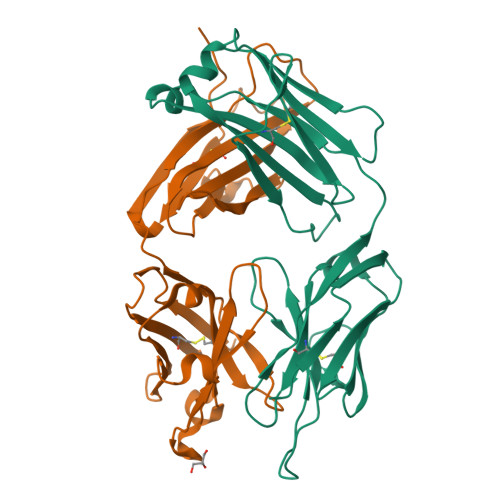
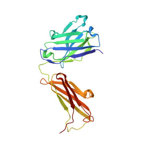
3I2C

Crystal structure of anti-IL-23 antibody CNTO4088


Experimental Data Snapshot

  • Method: X-RAY DIFFRACTION
  • Resolution: 2.80 Å
  • R-Value Free: 0.235 
  • R-Value Work: 0.196 
  • R-Value Observed: 0.198 

Starting Model: experimental
View more details

wwPDB Validation   3D Report Full Report


This is version 1.5 of the entry. See complete history


Literature

Noncanonical conformation of CDR L1 in the anti-IL-23 antibody CNTO4088.

Teplyakov, A.Obmolova, G.Rogers, A.Gilliland, G.L.

(2010) Acta Crystallogr Sect F Struct Biol Cryst Commun 66: 229-232

  • DOI: https://doi.org/10.1107/S1744309109054141
  • Primary Citation of Related Structures:  
    3I2C

  • PubMed Abstract: 

    CNTO4088 is a monoclonal antibody to human IL-23. The X-ray structure of the Fab fragment revealed an unusual noncanonical conformation of CDR L1. Most antibodies with the kappa light chain exhibit a canonical structure for CDR L1 in which residue 29 anchors the CDR loop to the framework. Analysis of the residues believed to define the conformation of CDR L1 did not explain why it should not adopt a canonical conformation in this antibody. This makes CNTO4088 a benchmark case for developing prediction methods and structure-modeling tools.


  • Organizational Affiliation

    Centocor R&D Inc., 145 King of Prussia Road, Radnor, PA 19087, USA. ateplyak@its.jnj.com


Macromolecules
Find similar proteins by:  (by identity cutoff)  |  3D Structure
Entity ID: 1
MoleculeChains Sequence LengthOrganismDetailsImage
CNTO4088 LIGHT CHAINA [auth L]218Mus musculusMutation(s): 0 
Entity Groups  
Sequence Clusters30% Identity50% Identity70% Identity90% Identity95% Identity100% Identity
Sequence Annotations
Expand
  • Reference Sequence
Find similar proteins by:  (by identity cutoff)  |  3D Structure
Entity ID: 2
MoleculeChains Sequence LengthOrganismDetailsImage
CNTO4088 HEAVY CHAINB [auth H]222Mus musculusMutation(s): 0 
Entity Groups  
Sequence Clusters30% Identity50% Identity70% Identity90% Identity95% Identity100% Identity
Sequence Annotations
Expand
  • Reference Sequence
Small Molecules
Ligands 1 Unique
IDChains Name / Formula / InChI Key2D Diagram3D Interactions
GOL
Query on GOL

Download Ideal Coordinates CCD File 
C [auth H]GLYCEROL
C3 H8 O3
PEDCQBHIVMGVHV-UHFFFAOYSA-N
Experimental Data & Validation

Experimental Data

  • Method: X-RAY DIFFRACTION
  • Resolution: 2.80 Å
  • R-Value Free: 0.235 
  • R-Value Work: 0.196 
  • R-Value Observed: 0.198 
  • Space Group: I 4
Unit Cell:
Length ( Å )Angle ( ˚ )
a = 99.43α = 90
b = 99.43β = 90
c = 115.04γ = 90
Software Package:
Software NamePurpose
AMoREphasing
REFMACrefinement
d*TREKdata reduction
d*TREKdata scaling

Structure Validation

View Full Validation Report


Entry History 

Deposition Data

Revision History  (Full details and data files)

  • Version 1.0: 2009-07-07
    Type: Initial release
  • Version 1.1: 2011-07-13
    Changes: Non-polymer description, Version format compliance
  • Version 1.2: 2012-03-21
    Changes: Database references
  • Version 1.3: 2017-11-01
    Changes: Refinement description
  • Version 1.4: 2023-09-06
    Changes: Data collection, Database references, Derived calculations, Refinement description
  • Version 1.5: 2024-10-09
    Changes: Structure summary