


Crystal structure of the RabGAP domain of the RABGAP1L protein
Nedyalkova, L., Tempel, W., Tong, Y., Zhong, N., MacKenzie, F., Arrowsmith, C.H., Edwards, A.M., Bountra, C., Weigelt, J., Bochkarev, A., Park, H.To be published.
Experimental Data Snapshot
wwPDB Validation 3D Report Full Report

Entity ID: 1 | |||||
|---|---|---|---|---|---|
| Molecule | Chains | Sequence Length | Organism | Details | Image |
| RAB GTPase-activating protein 1-like | 310 | Homo sapiens | Mutation(s): 0 Gene Names: RABGAP1L, HHL, KIAA0471 | 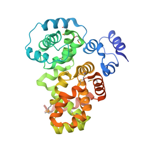 | |
UniProt & NIH Common Fund Data Resources | |||||
PHAROS: Q5R372 GTEx: ENSG00000152061 | |||||
Entity Groups | |||||
| Sequence Clusters | 30% Identity50% Identity70% Identity90% Identity95% Identity100% Identity | ||||
| UniProt Group | Q5R372 | ||||
Sequence AnnotationsExpand | |||||
Reference Sequence | |||||
| Ligands 1 Unique | |||||
|---|---|---|---|---|---|
| ID | Chains | Name / Formula / InChI Key | 2D Diagram | 3D Interactions | |
| UNX Download:Ideal Coordinates CCD File | AA [auth C] BA [auth C] CA [auth C] D [auth A] DA [auth C] | UNKNOWN ATOM OR ION X |  | ||
| Modified Residues 1 Unique | |||||
|---|---|---|---|---|---|
| ID | Chains | Type | Formula | 2D Diagram | Parent |
| MSE Query on MSE | A, B, C | L-PEPTIDE LINKING | C5 H11 N O2 Se |  | MET |
| Length ( Å ) | Angle ( ˚ ) |
|---|---|
| a = 48.086 | α = 90 |
| b = 64.571 | β = 90 |
| c = 290.255 | γ = 90 |
| Software Name | Purpose |
|---|---|
| DENZO | data reduction |
| SCALEPACK | data scaling |
| SHELX | phasing |
| RESOLVE | phasing |
| REFMAC | refinement |
| PDB_EXTRACT | data extraction |
| HKL-2000 | data reduction |
| HKL-2000 | data scaling |


