3HY7

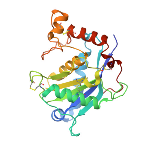
Crystal Structure of the Catalytic Domain of ADAMTS-5 in Complex with Marimastat


Experimental Data Snapshot

  • Method: X-RAY DIFFRACTION
  • Resolution: 1.69 Å
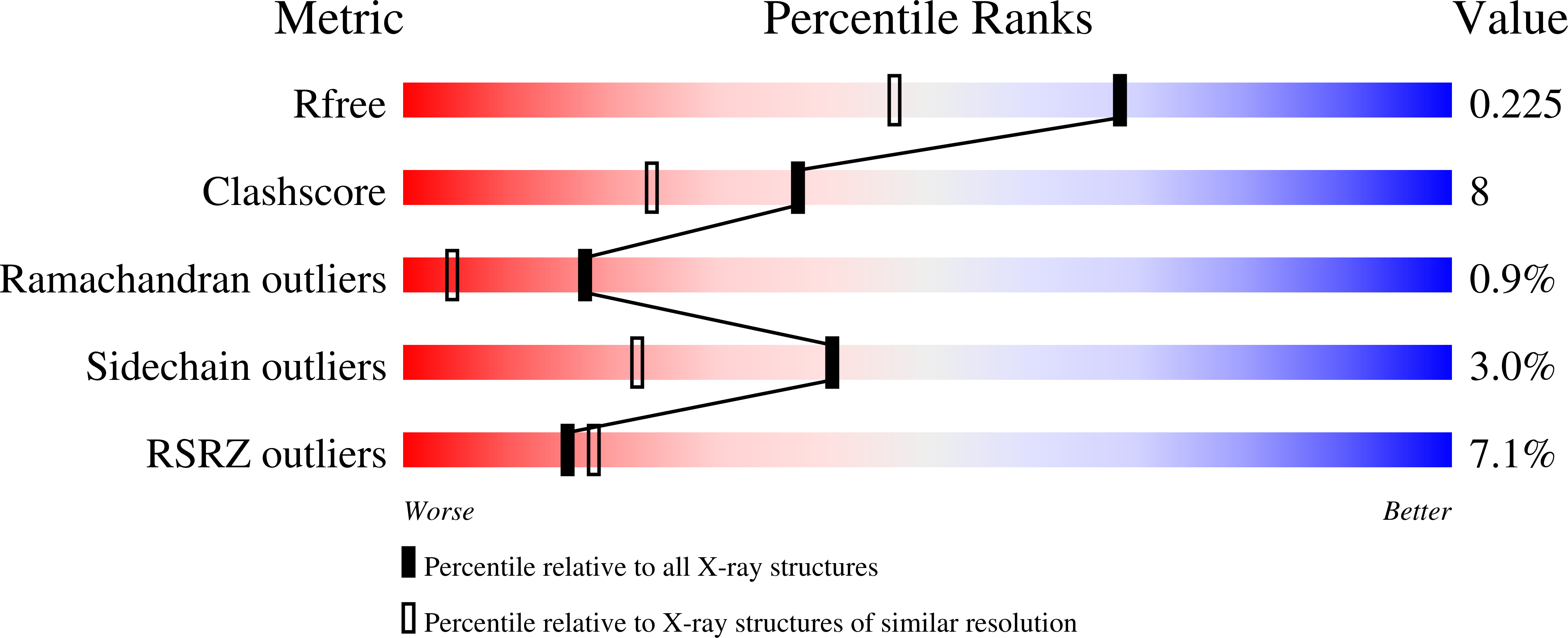
  • R-Value Free: 0.224 
  • R-Value Work: 0.173 
  • R-Value Observed: 0.176 

Starting Model: experimental
View more details

wwPDB Validation   3D Report Full Report


Ligand Structure Quality Assessment 


This is version 1.4 of the entry. See complete history


Literature

Structural and inhibition analysis reveals the mechanism of selectivity of a series of aggrecanase inhibitors

Tortorella, M.D.Tomasselli, A.G.Mathis, K.J.Schnute, M.E.Woodard, S.S.Munie, G.Williams, J.M.Caspers, N.Wittwer, A.J.Malfait, A.M.Shieh, H.S.

(2009) J Biol Chem 284: 24185-24191

  • DOI: https://doi.org/10.1074/jbc.M109.029116
  • Primary Citation of Related Structures:  
    3HY7, 3HY9, 3HYG

  • PubMed Abstract: 

    Several inhibitors of a series of cis-1(S)2(R)-amino-2-indanol-based compounds were reported to be selective for the aggrecanases, ADAMTS-4 and -5 over other metalloproteases. To understand the nature of this selectivity for aggrecanases, the inhibitors, along with the broad spectrum metalloprotease inhibitor marimastat, were independently bound to the catalytic domain of ADAMTS-5, and the corresponding crystal structures were determined. By comparing the structures, it was determined that the specificity of the relative inhibitors for ADAMTS-5 was not driven by a specific interaction, such as zinc chelation, hydrogen bonding, or charge interactions, but rather by subtle and indirect factors, such as water bridging, ring rigidity, pocket size, and shape, as well as protein conformation flexibility.


  • Organizational Affiliation

    Pfizer Global Research and Development, St. Louis, Missouri 63017, USA. micky.d.tortorella@gmail.com


Macromolecules
Find similar proteins by:  (by identity cutoff)  |  3D Structure
Entity ID: 1
MoleculeChains Sequence LengthOrganismDetailsImage
A disintegrin and metalloproteinase with thrombospondin motifs 5
A, B
221Homo sapiensMutation(s): 1 
Gene Names: ADAMTS-5ADAMTS11ADAMTS5ADMP2
EC: 3.4.24
UniProt & NIH Common Fund Data Resources
Find proteins for Q9UNA0 (Homo sapiens)
Go to UniProtKB:  Q9UNA0
PHAROS:  Q9UNA0
GTEx:  ENSG00000154736 
Sequence Annotations
Expand
  • Reference Sequence
Small Molecules
Ligands 3 Unique
IDChains Name / Formula / InChI Key2D Diagram3D Interactions
097
Query on 097

Download Ideal Coordinates CCD File 
C [auth A],
H [auth B]
(2S,3R)-N~4~-[(1S)-2,2-dimethyl-1-(methylcarbamoyl)propyl]-N~1~,2-dihydroxy-3-(2-methylpropyl)butanediamide
C15 H29 N3 O5
OCSMOTCMPXTDND-OUAUKWLOSA-N
ZN
Query on ZN

Download Ideal Coordinates CCD File 
D [auth A],
I [auth B]
ZINC ION
Zn
PTFCDOFLOPIGGS-UHFFFAOYSA-N
CA
Query on CA

Download Ideal Coordinates CCD File 
E [auth A]
F [auth A]
G [auth A]
J [auth B]
K [auth B]
E [auth A],
F [auth A],
G [auth A],
J [auth B],
K [auth B],
L [auth B]
CALCIUM ION
Ca
BHPQYMZQTOCNFJ-UHFFFAOYSA-N
Binding Affinity Annotations 
IDSourceBinding Affinity
097 BindingDB:  3HY7 IC50: 1.00e+4 (nM) from 1 assay(s)
Experimental Data & Validation

Experimental Data

  • Method: X-RAY DIFFRACTION
  • Resolution: 1.69 Å
  • R-Value Free: 0.224 
  • R-Value Work: 0.173 
  • R-Value Observed: 0.176 
  • Space Group: P 1 21 1
Unit Cell:
Length ( Å )Angle ( ˚ )
a = 52.606α = 90
b = 44.464β = 89.81
c = 76.349γ = 90
Software Package:
Software NamePurpose
HKL-2000data collection
REFMACrefinement
HKL-2000data reduction
HKL-2000data scaling

Structure Validation

View Full Validation Report


Ligand Structure Quality Assessment 


Entry History 

Deposition Data

Revision History  (Full details and data files)

  • Version 1.0: 2009-07-07
    Type: Initial release
  • Version 1.1: 2011-07-13
    Changes: Version format compliance
  • Version 1.2: 2017-11-01
    Changes: Refinement description
  • Version 1.3: 2021-10-13
    Changes: Database references, Derived calculations
  • Version 1.4: 2023-09-06
    Changes: Data collection, Refinement description