3HWS | pdb_00003hws

Crystal structure of nucleotide-bound hexameric ClpX


Experimental Data Snapshot

  • Method: X-RAY DIFFRACTION
  • Resolution: 3.25 Å
  • R-Value Free: 
    0.282 (Depositor), 0.283 (DCC) 
  • R-Value Work: 
    0.243 (Depositor), 0.241 (DCC) 
  • R-Value Observed: 
    0.245 (Depositor) 

wwPDB Validation 3D Report Full Report

Validation slider image for 3HWS

Ligand Structure Quality Assessment 


This is version 1.5 of the entry. See complete history

Literature

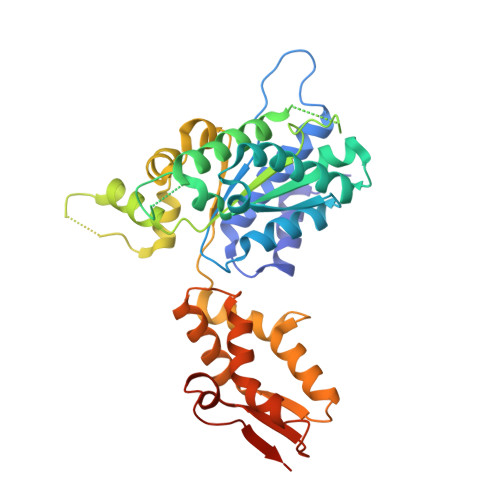
Structures of asymmetric ClpX hexamers reveal nucleotide-dependent motions in a AAA+ protein-unfolding machine.

Glynn, S.E.Martin, A.Nager, A.R.Baker, T.A.Sauer, R.T.

(2009) Cell 139: 744-756

  • DOI: https://doi.org/10.1016/j.cell.2009.09.034
  • Primary Citation Related Structures: 
    3HTE, 3HWS

  • PubMed Abstract: 

    ClpX is a AAA+ machine that uses the energy of ATP binding and hydrolysis to unfold native proteins and translocate unfolded polypeptides into the ClpP peptidase. The crystal structures presented here reveal striking asymmetry in ring hexamers of nucleotide-free and nucleotide-bound ClpX. Asymmetry arises from large changes in rotation between the large and small AAA+ domains of individual subunits. These differences prevent nucleotide binding to two subunits, generate a staggered arrangement of ClpX subunits and pore loops around the hexameric ring, and provide a mechanism for coupling conformational changes caused by ATP binding or hydrolysis in one subunit to flexing motions of the entire ring. Our structures explain numerous solution studies of ClpX function, predict mechanisms for pore elasticity during translocation of irregular polypeptides, and suggest how repetitive conformational changes might be coupled to mechanical work during the ATPase cycle of ClpX and related molecular machines.


  • Organizational Affiliation
    • Department of Biology, Howard Hughes Medical Institute, Massachusetts Institute of Technology, Cambridge, MA 02139, USA.

Macromolecule Content 

  • Total Structure Weight: 239.62 kDa 
  • Atom Count: 14,292 
  • Modeled Residue Count: 1,885 
  • Deposited Residue Count: 2,178 
  • Unique protein chains: 1

Macromolecules

Find similar proteins by:|  3D Structure
Entity ID: 1
MoleculeChains  Sequence LengthOrganismDetailsImage
ATP-dependent Clp protease ATP-binding subunit clpX
A, B, C, D, E
A, B, C, D, E, F
363Escherichia coli K-12Mutation(s): 2 
Gene Names: b0438clpXJW0428lopC
UniProt
Find proteins for P0A6H1 (Escherichia coli (strain K12))
Explore P0A6H1 
Go to UniProtKB:  P0A6H1
Entity Groups
Sequence Clusters30% Identity50% Identity70% Identity90% Identity95% Identity100% Identity
UniProt GroupP0A6H1
Sequence Annotations
Expand
Reference Sequence

Small Molecules

Ligands 3 Unique
IDChains Name / Formula / InChI Key2D Diagram3D Interactions
ADP

Query on ADP


Download:Ideal Coordinates CCD File
G [auth A],
K [auth B],
U [auth D],
X [auth E]
ADENOSINE-5'-DIPHOSPHATE
C10 H15 N5 O10 P2
XTWYTFMLZFPYCI-KQYNXXCUSA-N
SO4

Query on SO4


Download:Ideal Coordinates CCD File
H [auth A]
I [auth A]
J [auth A]
L [auth B]
M [auth B]
H [auth A],
I [auth A],
J [auth A],
L [auth B],
M [auth B],
N [auth B],
O [auth B],
Q [auth C],
R [auth C],
S [auth C],
T [auth C],
V [auth D],
Y [auth F]
SULFATE ION
O4 S
QAOWNCQODCNURD-UHFFFAOYSA-L
MG

Query on MG


Download:Ideal Coordinates CCD File
P [auth B],
W [auth D]
MAGNESIUM ION
Mg
JLVVSXFLKOJNIY-UHFFFAOYSA-N

Experimental Data & Validation

Experimental Data

  • Method: X-RAY DIFFRACTION
  • Resolution: 3.25 Å
  • R-Value Free:  0.282 (Depositor), 0.283 (DCC) 
  • R-Value Work:  0.243 (Depositor), 0.241 (DCC) 
  • R-Value Observed: 0.245 (Depositor) 
Space Group: P 21 21 21
Unit Cell:
Length ( Å )Angle ( ˚ )
a = 54.085α = 90
b = 178.498β = 90
c = 201.369γ = 90
Software Package:
Software NamePurpose
DENZOdata reduction
SCALEPACKdata scaling
PHENIXrefinement
PDB_EXTRACTdata extraction
ADSCdata collection
HKL-2000data reduction
HKL-2000data scaling
PHASERphasing

Structure Validation

View Full Validation Report


Ligand Structure Quality Assessment 


Entry History 

Deposition Data

Revision History  (Full details and data files)

  • Version 1.0: 2009-11-24
    Type: Initial release
  • Version 1.1: 2011-07-13
    Changes: Source and taxonomy, Version format compliance
  • Version 1.2: 2012-03-21
    Changes: Database references
  • Version 1.3: 2017-11-01
    Changes: Advisory, Refinement description
  • Version 1.4: 2021-10-13
    Changes: Advisory, Database references, Derived calculations
  • Version 1.5: 2024-02-21
    Changes: Data collection