3HWN | pdb_00003hwn

CATHEPSIN L with AZ13010160


Experimental Data Snapshot

  • Method: X-RAY DIFFRACTION
  • Resolution: 2.33 Å
  • R-Value Free: 
    0.347 (Depositor), 0.358 (DCC) 
  • R-Value Work: 
    0.250 (Depositor), 0.258 (DCC) 
  • R-Value Observed: 
    0.252 (Depositor) 

wwPDB Validation 3D Report Full Report

Validation slider image for 3HWN

Ligand Structure Quality Assessment 


This is version 1.4 of the entry. See complete history

Literature

Design of selective Cathepsin inhibitors

Bethel, P.A.Gerhardt, S.Jones, E.V.Kenny, P.W.Karoutchi, G.I.Morley, A.D.Oldham, K.Rankine, N.Augustin, M.Krapp, S.Simader, H.Steinbacher, S.

(2009) Bioorg Med Chem Lett 19: 4622-4625

  • DOI: https://doi.org/10.1016/j.bmcl.2009.06.090
  • Primary Citation Related Structures: 
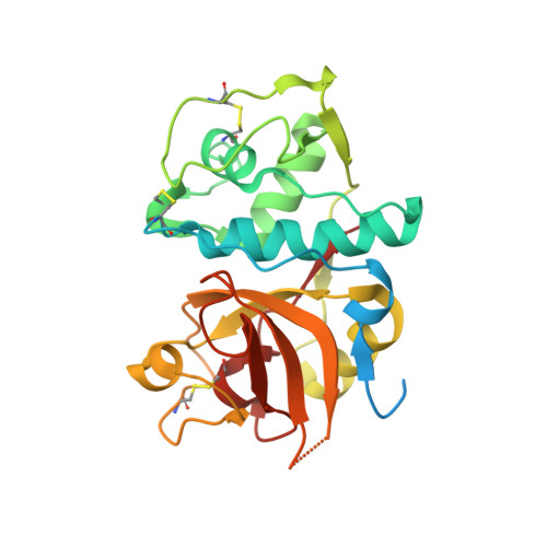
    3HWN

  • PubMed Abstract: 

    A number of molecular recognition features have been exploited in structure-based design of selective Cathepsin inhibitors.


  • Organizational Affiliation
    • RIRA, AstraZeneca, Mereside, Alderley Park, Macclesfield SK10 4TG, UK.

Macromolecule Content 

  • Total Structure Weight: 116.57 kDa 
  • Atom Count: 6,934 
  • Modeled Residue Count: 862 
  • Deposited Residue Count: 1,032 
  • Unique protein chains: 1

Macromolecules

Find similar proteins by:|  3D Structure
Entity ID: 1
MoleculeChains  Sequence LengthOrganismDetailsImage
Cathepsin L1
A, B, C, D
258Homo sapiensMutation(s): 1 
Gene Names: CTSL1CTSL
EC: 3.4.22.15
UniProt & NIH Common Fund Data Resources
Find proteins for P07711 (Homo sapiens)
Explore P07711 
Go to UniProtKB:  P07711
PHAROS:  P07711
GTEx:  ENSG00000135047 
Entity Groups
Sequence Clusters30% Identity50% Identity70% Identity90% Identity95% Identity100% Identity
UniProt GroupP07711
Sequence Annotations
Expand
Reference Sequence

Experimental Data & Validation

Experimental Data

  • Method: X-RAY DIFFRACTION
  • Resolution: 2.33 Å
  • R-Value Free:  0.347 (Depositor), 0.358 (DCC) 
  • R-Value Work:  0.250 (Depositor), 0.258 (DCC) 
  • R-Value Observed: 0.252 (Depositor) 
Space Group: P 1
Unit Cell:
Length ( Å )Angle ( ˚ )
a = 57.07α = 105.34
b = 62.8β = 94.04
c = 67.97γ = 115.26
Software Package:
Software NamePurpose
MOLREPphasing
REFMACrefinement
XDSdata reduction
XSCALEdata scaling

Structure Validation

View Full Validation Report


Ligand Structure Quality Assessment 


Entry History 

Deposition Data

Revision History  (Full details and data files)

  • Version 1.0: 2009-09-15
    Type: Initial release
  • Version 1.1: 2011-07-13
    Changes: Version format compliance
  • Version 1.2: 2017-11-01
    Changes: Advisory, Refinement description
  • Version 1.3: 2021-10-13
    Changes: Advisory, Database references, Derived calculations
  • Version 1.4: 2024-11-06
    Changes: Data collection, Structure summary