3HW7 | pdb_00003hw7

High pressure (0.57 GPa) crystal structure of bovine copper, zinc superoxide dismutase at 2.0 angstroms


Experimental Data Snapshot

  • Method: X-RAY DIFFRACTION
  • Resolution: 2.00 Å
  • R-Value Free: 
    0.218 (Depositor), 0.210 (DCC) 
  • R-Value Work: 
    0.172 (Depositor) 
  • R-Value Observed: 
    0.174 (Depositor) 

Starting Model: experimental
View more details

wwPDB Validation 3D Report Full Report

Validation slider image for 3HW7

This is version 1.4 of the entry. See complete history

Literature

Flexibility of the Cu,Zn superoxide dismutase structure investigated at 0.57 GPa

Ascone, I.Savino, C.Kahn, R.Fourme, R.

(2010) Acta Crystallogr D Biol Crystallogr 66: 654-663

  • DOI: https://doi.org/10.1107/S0907444910012321
  • Primary Citation Related Structures: 
    3HW7

  • PubMed Abstract: 

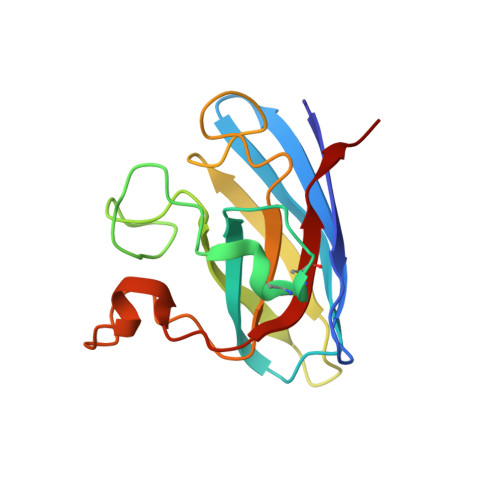
    The 2 A resolution crystal structure of bovine erythrocyte Cu,Zn superoxide dismutase (CuZnSOD) has been determined by X-ray diffraction at high pressure (0.57 GPa) and room temperature. At 0.57 GPa the secondary, tertiary and quaternary structures are similar to other previously determined bovine erythrocyte CuZnSOD structures. Nevertheless, pressure has a localized impact on the atomic coordinates of C(alpha) atoms and on side chains. The compression of the crystal and of the protein backbone is anisotropic. This anisotropy is discussed, taking into account intermolecular contacts and protein conformation. Pressure perturbation highlights the more flexible zones in the protein such as the electrostatic loop. At 0.57 GPa, a global shift of the dimetallic sites in both subunits and changes in the oxidation state of Cu were observed. The flexibility of the electrostatic loop may be useful for the interaction of different metal carriers in the copper-uptake process, whereas the flexibility of the metal sites involved in the activity of the protein could contribute to explaining the ubiquitous character of CuZnSODs, which are found in organisms living in very different conditions, including the deep-sea environment. This work illustrates the potential of combining X-ray crystallography with high pressure to promote and stabilize higher energy conformational substates.


  • Organizational Affiliation
    • ENSCP, UMR CNRS 7223, 11 Rue Pierre et Marie Curie, 75231 Paris CEDEX 05, France. gchojnowski@iimcb.gov.pl

Macromolecule Content 

  • Total Structure Weight: 31.47 kDa 
  • Atom Count: 2,395 
  • Modeled Residue Count: 302 
  • Deposited Residue Count: 302 
  • Unique protein chains: 1

Macromolecules

Find similar proteins by:|  3D Structure
Entity ID: 1
MoleculeChains  Sequence LengthOrganismDetailsImage
Superoxide dismutase [Cu-Zn]
A, B
151Bos taurusMutation(s): 0 
EC: 1.15.1.1 (PDB Primary Data), 1.8 (UniProt)
UniProt
Find proteins for P00442 (Bos taurus)
Explore P00442 
Go to UniProtKB:  P00442
Entity Groups
Sequence Clusters30% Identity50% Identity70% Identity90% Identity95% Identity100% Identity
UniProt GroupP00442
Sequence Annotations
Expand
Reference Sequence

Experimental Data & Validation

Experimental Data

  • Method: X-RAY DIFFRACTION
  • Resolution: 2.00 Å
  • R-Value Free:  0.218 (Depositor), 0.210 (DCC) 
  • R-Value Work:  0.172 (Depositor) 
  • R-Value Observed: 0.174 (Depositor) 
Space Group: P 21 21 21
Unit Cell:
Length ( Å )Angle ( ˚ )
a = 46.845α = 90
b = 50.509β = 90
c = 146.377γ = 90
Software Package:
Software NamePurpose
MAR345dtbdata collection
CCP4model building
REFMACrefinement
XDSdata reduction
SCALAdata scaling
CCP4phasing

Structure Validation

View Full Validation Report


Entry History 

Deposition Data

Revision History  (Full details and data files)

  • Version 1.0: 2010-06-23
    Type: Initial release
  • Version 1.1: 2011-07-13
    Changes: Version format compliance
  • Version 1.2: 2012-04-18
    Changes: Database references
  • Version 1.3: 2023-11-01
    Changes: Data collection, Database references, Derived calculations, Refinement description
  • Version 1.4: 2024-11-20
    Changes: Structure summary