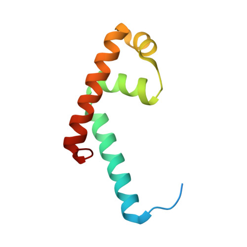
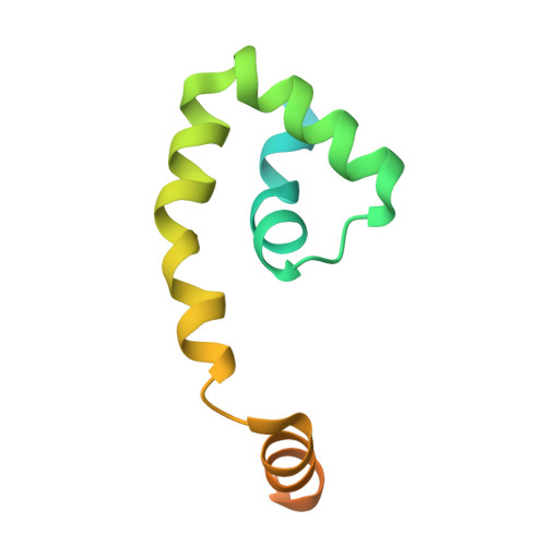
3HUG

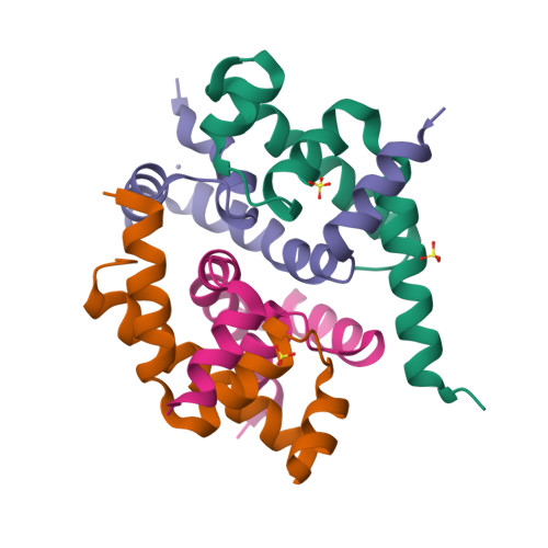
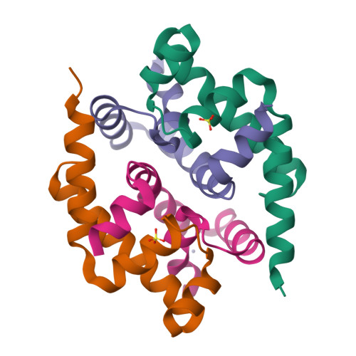
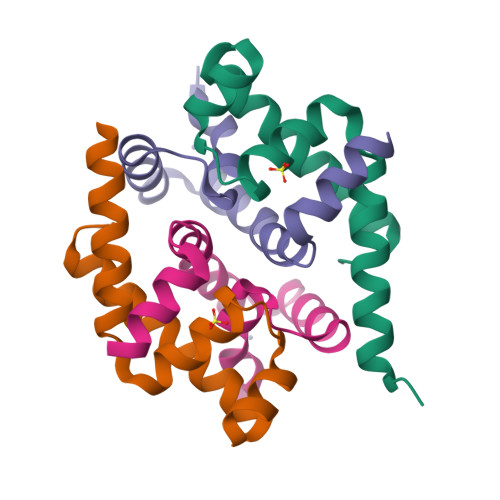
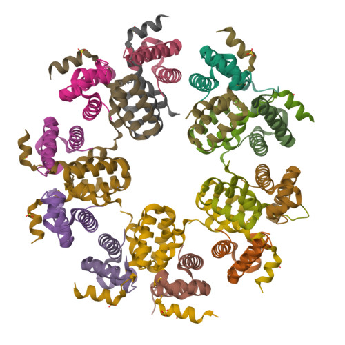
Crystal structure of Mycobacterium tuberculosis anti-sigma factor RslA in complex with -35 promoter binding domain of sigL


Experimental Data Snapshot

  • Method: X-RAY DIFFRACTION
  • Resolution: 2.35 Å
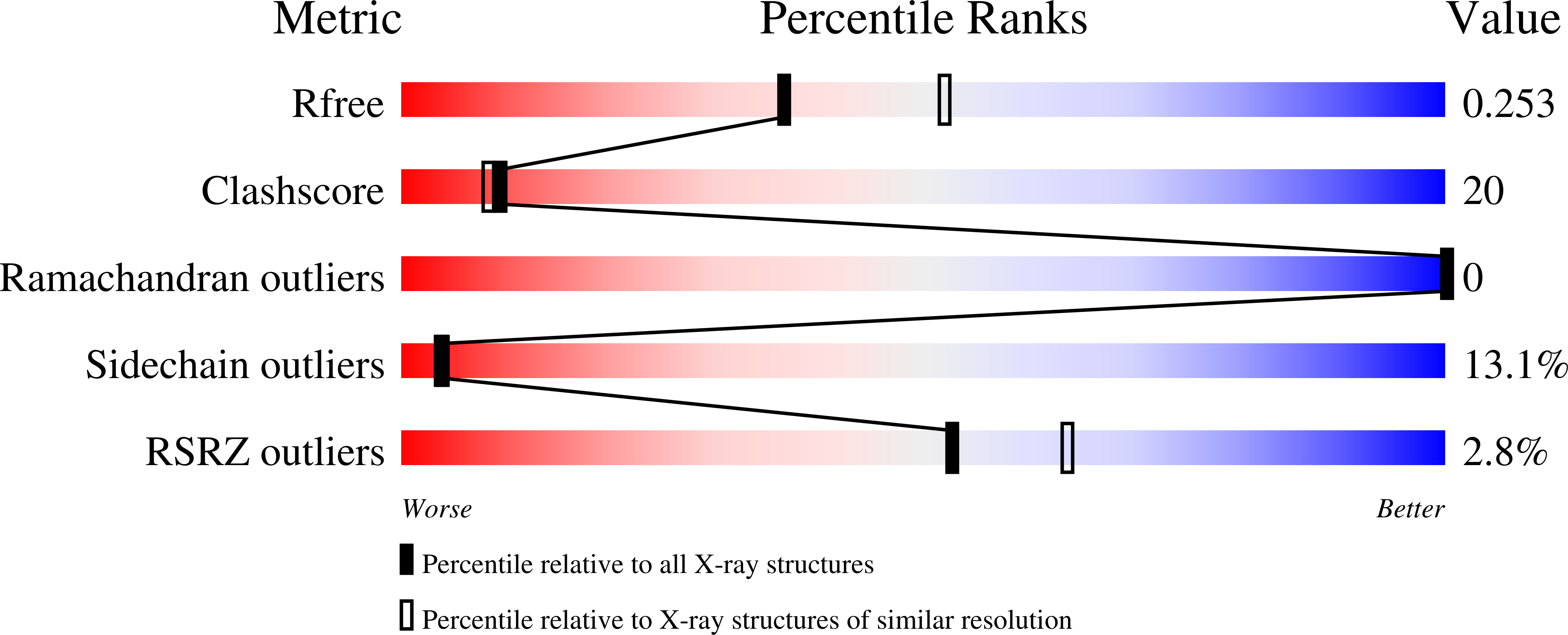
  • R-Value Free: 0.260 
  • R-Value Work: 0.211 
  • R-Value Observed: 0.213 

wwPDB Validation   3D Report Full Report


This is version 1.3 of the entry. See complete history


Literature

Structural and biochemical bases for the redox sensitivity of Mycobacterium tuberculosis RslA

Thakur, K.G.Praveena, T.Gopal, B.

(2010) J Mol Biol 397: 1199-1208

  • DOI: https://doi.org/10.1016/j.jmb.2010.02.026
  • Primary Citation of Related Structures:  
    3HUG

  • PubMed Abstract: 

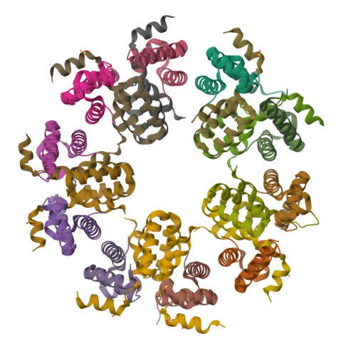
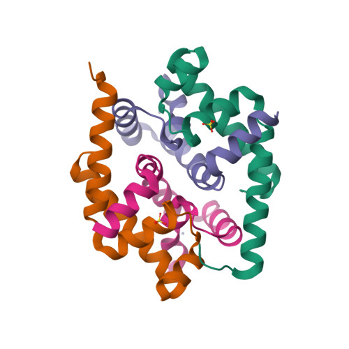
    An effective transcriptional response to redox stimuli is of particular importance for Mycobacterium tuberculosis, as it adapts to the environment of host alveoli and macrophages. The M. tuberculosis sigma factor sigma(L) regulates the expression of genes involved in cell-wall and polyketide syntheses. sigma(L) interacts with the cytosolic anti-sigma domain of a membrane-associated protein, RslA. Here we demonstrate that RslA binds Zn(2+) and can sequester sigma(L) in a reducing environment. In response to an oxidative stimulus, proximal cysteines in the CXXC motif of RslA form a disulfide bond, releasing bound Zn(2+). This results in a substantial rearrangement of the sigma(L)/RslA complex, leading to an 8-fold decrease in the affinity of RslA for sigma(L). The crystal structure of the -35-element recognition domain of sigma(L), sigma(4)(L), bound to RslA reveals that RslA inactivates sigma(L) by sterically occluding promoter DNA and RNA polymerase binding sites. The crystal structure further reveals that the cysteine residues that coordinate Zn(2+) in RslA are solvent exposed in the complex, thus providing a structural basis for the redox sensitivity of RslA. The biophysical parameters of sigma(L)/RslA interactions provide a template for understanding how variations in the rate of Zn(2+) release and associated conformational changes could regulate the activity of a Zn(2+)-associated anti-sigma factor.


  • Organizational Affiliation

    Molecular Biophysics Unit, Indian Institute of Science, Bangalore 560 012, India.


Macromolecules
Find similar proteins by:  (by identity cutoff)  |  3D Structure
Entity ID: 1
MoleculeChains Sequence LengthOrganismDetailsImage
RNA polymerase sigma factor
A, C, E, G, I
A, C, E, G, I, K, M, O, Q, S
92Mycobacterium tuberculosis H37RvMutation(s): 0 
Gene Names: sigL
UniProt
Find proteins for P9WGH5 (Mycobacterium tuberculosis (strain ATCC 25618 / H37Rv))
Explore P9WGH5 
Go to UniProtKB:  P9WGH5
Entity Groups  
Sequence Clusters30% Identity50% Identity70% Identity90% Identity95% Identity100% Identity
UniProt GroupP9WGH5
Sequence Annotations
Expand
  • Reference Sequence
Find similar proteins by:  (by identity cutoff)  |  3D Structure
Entity ID: 2
MoleculeChains Sequence LengthOrganismDetailsImage
PROBABLE CONSERVED MEMBRANE PROTEIN
B, D, F, H, J
B, D, F, H, J, L, N, P, R, T
108Mycobacterium tuberculosis H37RvMutation(s): 0 
Gene Names: RslA (Rv0736)
UniProt
Find proteins for P9WJ67 (Mycobacterium tuberculosis (strain ATCC 25618 / H37Rv))
Explore P9WJ67 
Go to UniProtKB:  P9WJ67
Entity Groups  
Sequence Clusters30% Identity50% Identity70% Identity90% Identity95% Identity100% Identity
UniProt GroupP9WJ67
Sequence Annotations
Expand
  • Reference Sequence
Small Molecules
Ligands 2 Unique
IDChains Name / Formula / InChI Key2D Diagram3D Interactions
SO4
Query on SO4

Download Ideal Coordinates CCD File 
BA [auth I]
CA [auth I]
EA [auth L]
GA [auth M]
IA [auth P]
BA [auth I],
CA [auth I],
EA [auth L],
GA [auth M],
IA [auth P],
KA [auth Q],
MA [auth S],
U [auth A],
W [auth C],
Z [auth G]
SULFATE ION
O4 S
QAOWNCQODCNURD-UHFFFAOYSA-L
ZN
Query on ZN

Download Ideal Coordinates CCD File 
AA [auth H]
DA [auth J]
FA [auth L]
HA [auth N]
JA [auth P]
AA [auth H],
DA [auth J],
FA [auth L],
HA [auth N],
JA [auth P],
LA [auth R],
NA [auth T],
V [auth B],
X [auth D],
Y [auth F]
ZINC ION
Zn
PTFCDOFLOPIGGS-UHFFFAOYSA-N
Experimental Data & Validation

Experimental Data

  • Method: X-RAY DIFFRACTION
  • Resolution: 2.35 Å
  • R-Value Free: 0.260 
  • R-Value Work: 0.211 
  • R-Value Observed: 0.213 
  • Space Group: P 21 21 21
Unit Cell:
Length ( Å )Angle ( ˚ )
a = 83.07α = 90
b = 166.77β = 90
c = 178.87γ = 90
Software Package:
Software NamePurpose
PHENIXmodel building
REFMACrefinement
MOSFLMdata reduction
SCALAdata scaling
PHENIXphasing

Structure Validation

View Full Validation Report


Entry History 

Deposition Data

Revision History  (Full details and data files)

  • Version 1.0: 2010-03-02
    Type: Initial release
  • Version 1.1: 2011-07-13
    Changes: Version format compliance
  • Version 1.2: 2011-09-28
    Changes: Database references, Refinement description
  • Version 1.3: 2024-03-20
    Changes: Data collection, Database references, Derived calculations