3HS4 | pdb_00003hs4

Human carbonic anhydrase II complexed with acetazolamide


Experimental Data Snapshot

  • Method: X-RAY DIFFRACTION
  • Resolution: 1.10 Å
  • R-Value Free: 
    0.140 (Depositor), 0.136 (DCC) 
  • R-Value Work: 
    0.112 (Depositor), 0.117 (DCC) 
  • R-Value Observed: 
    0.113 (Depositor) 

Starting Model: experimental
View more details

wwPDB Validation 3D Report Full Report

Validation slider image for 3HS4

Ligand Structure Quality Assessment 


This is version 1.3 of the entry. See complete history

Literature

High-resolution structure of human carbonic anhydrase II complexed with acetazolamide reveals insights into inhibitor drug design.

Sippel, K.H.Robbins, A.H.Domsic, J.Genis, C.Agbandje-McKenna, M.McKenna, R.

(2009) Acta Crystallogr Sect F Struct Biol Cryst Commun 65: 992-995

  • DOI: https://doi.org/10.1107/S1744309109036665
  • Primary Citation Related Structures: 
    3HS4

  • PubMed Abstract: 

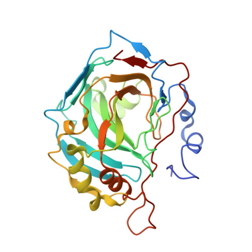
    The crystal structure of human carbonic anhydrase II (CA II) complexed with the inhibitor acetazolamide (AZM) has been determined at 1.1 A resolution and refined to an R(cryst) of 11.2% and an R(free) of 14.7%. As observed in previous CA II-inhibitor complexes, AZM binds directly to the zinc and makes several key interactions with active-site residues. The high-resolution data also showed a glycerol molecule adjacent to the AZM in the active site and two additional AZMs that are adventitiously bound on the surface of the enzyme. The co-binding of AZM and glycerol in the active site demonstrate that given an appropriate ring orientation and substituents, an isozyme-specific CA inhibitor may be developed.


  • Organizational Affiliation
    • Department of Biochemistry and Molecular Biology, College of Medicine, University of Florida, Gainesville, FL 32610, USA.

Macromolecule Content 

  • Total Structure Weight: 30.11 kDa 
  • Atom Count: 2,536 
  • Modeled Residue Count: 257 
  • Deposited Residue Count: 260 
  • Unique protein chains: 1

Macromolecules

Find similar proteins by:|  3D Structure
Entity ID: 1
MoleculeChains  Sequence LengthOrganismDetailsImage
Carbonic anhydrase 2260Homo sapiensMutation(s): 0 
Gene Names: CA2
EC: 4.2.1.1 (PDB Primary Data), 4.2.1.69 (UniProt)
UniProt & NIH Common Fund Data Resources
Find proteins for P00918 (Homo sapiens)
Explore P00918 
Go to UniProtKB:  P00918
PHAROS:  P00918
GTEx:  ENSG00000104267 
Entity Groups
Sequence Clusters30% Identity50% Identity70% Identity90% Identity95% Identity100% Identity
UniProt GroupP00918
Sequence Annotations
Expand
Reference Sequence

Experimental Data & Validation

Experimental Data

  • Method: X-RAY DIFFRACTION
  • Resolution: 1.10 Å
  • R-Value Free:  0.140 (Depositor), 0.136 (DCC) 
  • R-Value Work:  0.112 (Depositor), 0.117 (DCC) 
  • R-Value Observed: 0.113 (Depositor) 
Space Group: P 1 21 1
Unit Cell:
Length ( Å )Angle ( ˚ )
a = 42.252α = 90
b = 41.097β = 104.31
c = 71.82γ = 90
Software Package:
Software NamePurpose
SHELXL-97refinement
CNSrefinement
ADSCdata collection
HKL-2000data reduction
HKL-2000data scaling
CNSphasing

Structure Validation

View Full Validation Report


Ligand Structure Quality Assessment 


Entry History 

Deposition Data

Revision History  (Full details and data files)

  • Version 1.0: 2009-12-08
    Type: Initial release
  • Version 1.1: 2011-07-13
    Changes: Non-polymer description, Version format compliance
  • Version 1.2: 2019-07-24
    Changes: Data collection, Refinement description
  • Version 1.3: 2023-09-06
    Changes: Data collection, Database references, Derived calculations, Refinement description