3HQF | pdb_00003hqf

Crystal structure of restriction endonuclease EcoRII N-terminal effector-binding domain in complex with cognate DNA


Experimental Data Snapshot

  • Method: X-RAY DIFFRACTION
  • Resolution: 2.51 Å
  • R-Value Free: 
    0.229 (Depositor), 0.229 (DCC) 
  • R-Value Work: 
    0.187 (Depositor), 0.186 (DCC) 
  • R-Value Observed: 
    0.191 (Depositor) 

Starting Model: experimental
View more details

wwPDB Validation 3D Report Full Report

Validation slider image for 3HQF

This is version 1.2 of the entry. See complete history

Literature

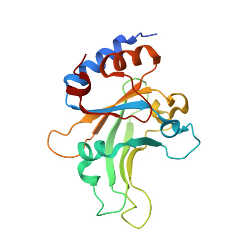
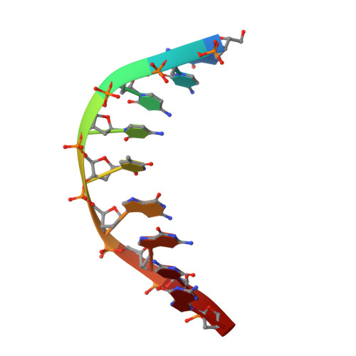
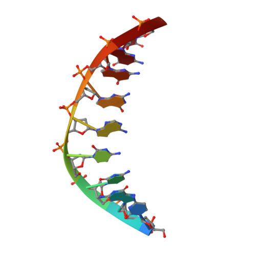
Structural mechanisms for the 5'-CCWGG sequence recognition by the N- and C-terminal domains of EcoRII.

Golovenko, D.Manakova, E.Tamulaitiene, G.Grazulis, S.Siksnys, V.

(2009) Nucleic Acids Res 37: 6613-6624

  • DOI: https://doi.org/10.1093/nar/gkp699
  • Primary Citation Related Structures: 
    3HQF, 3HQG

  • PubMed Abstract: 

    EcoRII restriction endonuclease is specific for the 5'-CCWGG sequence (W stands for A or T); however, it shows no activity on a single recognition site. To activate cleavage it requires binding of an additional target site as an allosteric effector. EcoRII dimer consists of three structural units: a central catalytic core, made from two copies of the C-terminal domain (EcoRII-C), and two N-terminal effector DNA binding domains (EcoRII-N). Here, we report DNA-bound EcoRII-N and EcoRII-C structures, which show that EcoRII combines two radically different structural mechanisms to interact with the effector and substrate DNA. The catalytic EcoRII-C dimer flips out the central T:A base pair and makes symmetric interactions with the CC:GG half-sites. The EcoRII-N effector domain monomer binds to the target site asymmetrically in a single defined orientation which is determined by specific hydrogen bonding and van der Waals interactions with the central T:A pair in the major groove. The EcoRII-N mode of the target site recognition is shared by the large class of higher plant transcription factors of the B3 superfamily.


  • Organizational Affiliation
    • Institute of Biotechnology, Graiciuno 8, LT-02241 Vilnius, Lithuania.

Macromolecule Content 

  • Total Structure Weight: 25.38 kDa 
  • Atom Count: 1,850 
  • Modeled Residue Count: 191 
  • Deposited Residue Count: 199 
  • Unique protein chains: 1
  • Unique nucleic acid chains: 2

Macromolecules


Find similar proteins by:|  3D Structure
Entity ID: 1
MoleculeChains  Sequence LengthOrganismDetailsImage
Restriction endonuclease181Escherichia coliMutation(s): 0 
Gene Names: EcoRII
EC: 3.1.21.4
UniProt
Find proteins for O69414 (Escherichia coli)
Explore O69414 
Go to UniProtKB:  O69414
Entity Groups
Sequence Clusters30% Identity50% Identity70% Identity90% Identity95% Identity100% Identity
UniProt GroupO69414
Sequence Annotations
Expand
Reference Sequence
Find similar nucleic acids by:  Sequence
Entity ID: 2
MoleculeChains LengthOrganismImage
5'-D(*GP*CP*CP*CP*TP*GP*GP*CP*G)-3'9N/A
Sequence Annotations
Expand
Reference Sequence
Find similar nucleic acids by:  Sequence
Entity ID: 3
MoleculeChains LengthOrganismImage
5'-D(*CP*GP*CP*CP*AP*GP*GP*GP*C)-3'9N/A
Sequence Annotations
Expand
Reference Sequence

Experimental Data & Validation

Experimental Data

  • Method: X-RAY DIFFRACTION
  • Resolution: 2.51 Å
  • R-Value Free:  0.229 (Depositor), 0.229 (DCC) 
  • R-Value Work:  0.187 (Depositor), 0.186 (DCC) 
  • R-Value Observed: 0.191 (Depositor) 
Space Group: P 43 21 2
Unit Cell:
Length ( Å )Angle ( ˚ )
a = 43.156α = 90
b = 43.156β = 90
c = 253.648γ = 90
Software Package:
Software NamePurpose
REFMACrefinement
AMoREphasing
MOSFLMdata reduction
SCALAdata scaling
PDB_EXTRACTdata extraction

Structure Validation

View Full Validation Report


Entry History 

Deposition Data

Revision History  (Full details and data files)

  • Version 1.0: 2009-09-22
    Type: Initial release
  • Version 1.1: 2011-07-13
    Changes: Advisory, Refinement description, Version format compliance
  • Version 1.2: 2023-09-06
    Changes: Data collection, Database references, Refinement description