


Probing the active site of M. tuberculosis LeuA
Koon, N., Squire, C.J., Baker, E.N.To be published.
Experimental Data Snapshot
Starting Model: experimental
View more details
wwPDB Validation 3D Report Full Report

Entity ID: 1 | |||||
|---|---|---|---|---|---|
| Molecule | Chains | Sequence Length | Organism | Details | Image |
| 2-isopropylmalate synthase | 644 | Mycobacterium tuberculosis H37Rv | Mutation(s): 0 Gene Names: leuA, MT3813, MTV025.058, Rv3710 EC: 2.3.3.13 | 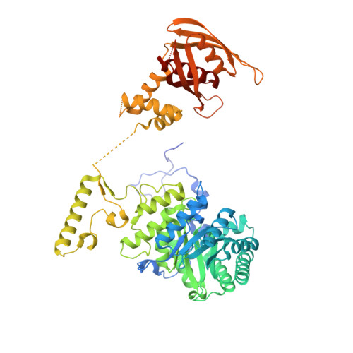 | |
UniProt | |||||
Entity Groups | |||||
| Sequence Clusters | 30% Identity50% Identity70% Identity90% Identity95% Identity100% Identity | ||||
| UniProt Group | P9WQB3 | ||||
Sequence AnnotationsExpand | |||||
Reference Sequence | |||||
| Ligands 4 Unique | |||||
|---|---|---|---|---|---|
| ID | Chains | Name / Formula / InChI Key | 2D Diagram | 3D Interactions | |
| LEU Download:Ideal Coordinates CCD File | C [auth A], G [auth B] | LEUCINE C6 H13 N O2 ROHFNLRQFUQHCH-YFKPBYRVSA-N |  | ||
| COI Download:Ideal Coordinates CCD File | E [auth A], I [auth B] | 2-OXO-4-METHYLPENTANOIC ACID C6 H10 O3 BKAJNAXTPSGJCU-UHFFFAOYSA-N |  | ||
| GOL Download:Ideal Coordinates CCD File | F [auth A], J [auth B] | GLYCEROL C3 H8 O3 PEDCQBHIVMGVHV-UHFFFAOYSA-N |  | ||
| ZN Download:Ideal Coordinates CCD File | D [auth A], H [auth B] | ZINC ION Zn PTFCDOFLOPIGGS-UHFFFAOYSA-N |  | ||
| Length ( Å ) | Angle ( ˚ ) |
|---|---|
| a = 54.411 | α = 90 |
| b = 154.848 | β = 97.65 |
| c = 68.882 | γ = 90 |
| Software Name | Purpose |
|---|---|
| MAR345dtb | data collection |
| REFMAC | refinement |
| MOSFLM | data reduction |
| SCALA | data scaling |
| REFMAC | phasing |


