3HNE | pdb_00003hne

Crystal structure of human ribonucleotide reductase 1 bound to the effectors TTP and ATP


Experimental Data Snapshot

  • Method: X-RAY DIFFRACTION
  • Resolution: 3.11 Å
  • R-Value Free: 
    0.275 (Depositor), 0.274 (DCC) 
  • R-Value Work: 
    0.185 (Depositor), 0.188 (DCC) 
  • R-Value Observed: 
    0.189 (Depositor) 

Starting Model: experimental
View more details

wwPDB Validation 3D Report Full Report

Validation slider image for 3HNE

Ligand Structure Quality Assessment 


This is version 1.3 of the entry. See complete history

Literature

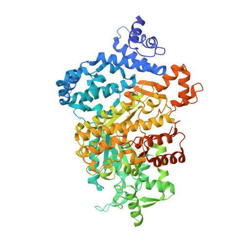
Structural basis for allosteric regulation of human ribonucleotide reductase by nucleotide-induced oligomerization.

Fairman, J.W.Wijerathna, S.R.Ahmad, M.F.Xu, H.Nakano, R.Jha, S.Prendergast, J.Welin, R.M.Flodin, S.Roos, A.Nordlund, P.Li, Z.Walz, T.Dealwis, C.G.

(2011) Nat Struct Mol Biol 18: 316-322

  • DOI: https://doi.org/10.1038/nsmb.2007
  • Primary Citation Related Structures: 
    2WGH, 3HNC, 3HND, 3HNE, 3HNF, 3PAW

  • PubMed Abstract: 

    Ribonucleotide reductase (RR) is an α(n)β(n) (RR1-RR2) complex that maintains balanced dNTP pools by reducing NDPs to dNDPs. RR1 is the catalytic subunit, and RR2 houses the free radical required for catalysis. RR is allosterically regulated by its activator ATP and its inhibitor dATP, which regulate RR activity by inducing oligomerization of RR1. Here, we report the first X-ray structures of human RR1 bound to TTP alone, dATP alone, TTP-GDP, TTP-ATP, and TTP-dATP. These structures provide insights into regulation of RR by ATP or dATP. At physiological dATP concentrations, RR1 forms inactive hexamers. We determined the first X-ray structure of the RR1-dATP hexamer and used single-particle electron microscopy to visualize the α(6)-ββ'-dATP holocomplex. Site-directed mutagenesis and functional assays confirm that hexamerization is a prerequisite for inhibition by dATP. Our data indicate a mechanism for regulating RR activity by dATP-induced oligomerization.


  • Organizational Affiliation
    • Department of Biochemistry, University of Tennessee, Knoxville, TN, USA.

Macromolecule Content 

  • Total Structure Weight: 182.5 kDa 
  • Atom Count: 11,380 
  • Modeled Residue Count: 1,439 
  • Deposited Residue Count: 1,584 
  • Unique protein chains: 1

Macromolecules

Find similar proteins by:|  3D Structure
Entity ID: 1
MoleculeChains  Sequence LengthOrganismDetailsImage
Ribonucleoside-diphosphate reductase large subunit
A, B
792Homo sapiensMutation(s): 0 
Gene Names: RR1RRM1
EC: 1.17.4.1
UniProt & NIH Common Fund Data Resources
Find proteins for P23921 (Homo sapiens)
Explore P23921 
Go to UniProtKB:  P23921
PHAROS:  P23921
GTEx:  ENSG00000167325 
Entity Groups
Sequence Clusters30% Identity50% Identity70% Identity90% Identity95% Identity100% Identity
UniProt GroupP23921
Sequence Annotations
Expand
Reference Sequence

Small Molecules

Ligands 4 Unique
IDChains Name / Formula / InChI Key2D Diagram3D Interactions
ATP

Query on ATP


Download:Ideal Coordinates CCD File
J [auth B]ADENOSINE-5'-TRIPHOSPHATE
C10 H16 N5 O13 P3
ZKHQWZAMYRWXGA-KQYNXXCUSA-N
TTP

Query on TTP


Download:Ideal Coordinates CCD File
C [auth A],
H [auth B]
THYMIDINE-5'-TRIPHOSPHATE
C10 H17 N2 O14 P3
NHVNXKFIZYSCEB-XLPZGREQSA-N
SO4

Query on SO4


Download:Ideal Coordinates CCD File
E [auth A]
F [auth A]
G [auth A]
M [auth B]
N [auth B]
E [auth A],
F [auth A],
G [auth A],
M [auth B],
N [auth B],
O [auth B]
SULFATE ION
O4 S
QAOWNCQODCNURD-UHFFFAOYSA-L
MG

Query on MG


Download:Ideal Coordinates CCD File
D [auth A],
I [auth B],
K [auth B],
L [auth B]
MAGNESIUM ION
Mg
JLVVSXFLKOJNIY-UHFFFAOYSA-N

Experimental Data & Validation

Experimental Data

  • Method: X-RAY DIFFRACTION
  • Resolution: 3.11 Å
  • R-Value Free:  0.275 (Depositor), 0.274 (DCC) 
  • R-Value Work:  0.185 (Depositor), 0.188 (DCC) 
  • R-Value Observed: 0.189 (Depositor) 
Space Group: P 21 21 21
Unit Cell:
Length ( Å )Angle ( ˚ )
a = 69.147α = 90
b = 114.372β = 90
c = 222.474γ = 90
Software Package:
Software NamePurpose
DENZOdata reduction
SCALEPACKdata scaling
PHASERphasing
REFMACrefinement
PDB_EXTRACTdata extraction
MAR345data collection

Structure Validation

View Full Validation Report


Ligand Structure Quality Assessment 


Entry History 

Deposition Data

Revision History  (Full details and data files)

  • Version 1.0: 2011-02-23
    Type: Initial release
  • Version 1.1: 2011-07-13
    Changes: Version format compliance
  • Version 1.2: 2017-11-01
    Changes: Advisory, Refinement description
  • Version 1.3: 2023-09-06
    Changes: Advisory, Data collection, Database references, Derived calculations, Refinement description