Identification of PDE4B Over 4D subtype-selective inhibitors revealing an unprecedented binding mode
Kranz, M., Wall, M., Evans, B., Miah, A., Ballantine, S., Delves, C., Dombroski, B., Gross, J., Schneck, J., Villa, J.P., Neu, M., Somers, D.O.(2009) Bioorg Med Chem 17: 5336-5341
- PubMed: 19525117 Search on PubMed
- DOI: https://doi.org/10.1016/j.bmc.2009.03.061
- Primary Citation Related Structures:
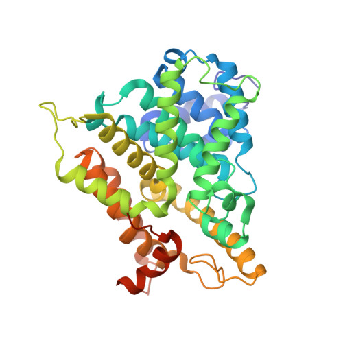
3HMV - PubMed Abstract:
A PDE4B over 4D-selective inhibitor programme was initiated to capitalise on the recently discovered predominance of the PDE4B subtype in inflammatory cell regulation. The SAR of a tetrahydrobenzothiophene (THBT) series did not agree with either of two proposed docking modes in the 4B binding site. A subsequent X-ray co-crystal structure determination revealed that the THBT ligand displaces the Gln-443 residue, invariably ligand-anchoring in previous PDE4 co-crystal structures, and even shifts helix-15 by 1-2A. For the first time, several residues of the C-terminus previously proposed to be involved in subtype selectivity are resolved and three of them extend into the ligand binding site potentially allowing for selective drug design.
- Medicines Research Centre, GlaxoSmithKline, Gunnels Wood Road, Stevenage, Hertfordshire, SG1 2NY, England, UK. michael.j.kranz@gsk.com
Organizational Affiliation: