3HLJ | pdb_00003hlj

Crystal structure of human carbonic anhydrase isozyme II with 3-methylthiobenzimidazo[1,2-c][1,2,3]thiadiazol-7-sulfonamide


Experimental Data Snapshot

  • Method: X-RAY DIFFRACTION
  • Resolution: 1.44 Å
  • R-Value Free: 
    0.206 (Depositor), 0.204 (DCC) 
  • R-Value Work: 
    0.175 (Depositor), 0.173 (DCC) 
  • R-Value Observed: 
    0.178 (Depositor) 

Starting Model: experimental
View more details

wwPDB Validation 3D Report Full Report

Validation slider image for 3HLJ

Ligand Structure Quality Assessment 


This is version 1.2 of the entry. See complete history

Literature

Inhibition and binding studies of carbonic anhydrase isozymes I, II and IX with benzimidazo[1,2-c][1,2,3]thiadiazole-7-sulphonamides

Baranauskiene, L.Hilvo, M.Matuliene, J.Golovenko, D.Manakova, E.Dudutiene, V.Michailoviene, V.Torresan, J.Jachno, J.Parkkila, S.Maresca, A.Supuran, C.T.Grazulis, S.Matulis, D.

(2010) J Enzyme Inhib Med Chem 25: 863-870

  • DOI: https://doi.org/10.3109/14756360903571685
  • Primary Citation Related Structures: 
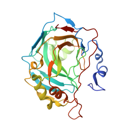
    3HLJ

  • PubMed Abstract: 

    The binding and inhibition strength of a series of benzimidazo[1,2-c][1,2,3]thiadiazole-7-sulphonamides were determined for recombinant human carbonic anhydrase isoforms I, II, and IX. The inhibition strength was determined by a stop-flow method to measure carbon dioxide hydration. Inhibitor-enzyme binding was determined by two biophysical techniques--isothermal titration calorimetry and thermal shift assay. The co-crystal structure was determined by X-ray crystallography. Comparing the results obtained using three different inhibition and binding methods increased the accuracy of compound affinity ranking and the ability to determine compound inhibitory specificity towards a particular carbonic anhydrase isoform. In most cases, all three methods yielded the same results despite using very different approaches to measure the binding and inhibition reactions. Some of the compounds studied are submicromolar inhibitors of the isoform IX, a prominent cancer target.


  • Organizational Affiliation
    • Laboratory of Biothermodynamics and Drug Design, Institute of Biotechnology, Graičiūno 8, Vilnius, Lithuania.

Macromolecule Content 

  • Total Structure Weight: 29.68 kDa 
  • Atom Count: 2,326 
  • Modeled Residue Count: 257 
  • Deposited Residue Count: 260 
  • Unique protein chains: 1

Macromolecules

Find similar proteins by:|  3D Structure
Entity ID: 1
MoleculeChains  Sequence LengthOrganismDetailsImage
Carbonic anhydrase 2260Homo sapiensMutation(s): 0 
Gene Names: CA2
EC: 4.2.1.1 (PDB Primary Data), 4.2.1.69 (UniProt)
UniProt & NIH Common Fund Data Resources
Find proteins for P00918 (Homo sapiens)
Explore P00918 
Go to UniProtKB:  P00918
PHAROS:  P00918
GTEx:  ENSG00000104267 
Entity Groups
Sequence Clusters30% Identity50% Identity70% Identity90% Identity95% Identity100% Identity
UniProt GroupP00918
Sequence Annotations
Expand
Reference Sequence

Small Molecules

Ligands 4 Unique
IDChains Name / Formula / InChI Key2D Diagram3D Interactions
V21

Query on V21


Download:Ideal Coordinates CCD File
B [auth A]3-methylthiobenzimidazo[1,2-c][1,2,3]thiadiazol-7-sulfonamide
C9 H8 N4 O2 S3
RQMLRBLXFGMTFD-UHFFFAOYSA-N
ZN

Query on ZN


Download:Ideal Coordinates CCD File
L [auth A]ZINC ION
Zn
PTFCDOFLOPIGGS-UHFFFAOYSA-N
NA

Query on NA


Download:Ideal Coordinates CCD File
C [auth A]SODIUM ION
Na
FKNQFGJONOIPTF-UHFFFAOYSA-N
UNX

Query on UNX


Download:Ideal Coordinates CCD File
D [auth A]
E [auth A]
F [auth A]
G [auth A]
H [auth A]
D [auth A],
E [auth A],
F [auth A],
G [auth A],
H [auth A],
I [auth A],
J [auth A],
K [auth A]
UNKNOWN ATOM OR ION
X

Experimental Data & Validation

Experimental Data

  • Method: X-RAY DIFFRACTION
  • Resolution: 1.44 Å
  • R-Value Free:  0.206 (Depositor), 0.204 (DCC) 
  • R-Value Work:  0.175 (Depositor), 0.173 (DCC) 
  • R-Value Observed: 0.178 (Depositor) 
Space Group: P 1 21 1
Unit Cell:
Length ( Å )Angle ( ˚ )
a = 42.14α = 90
b = 40.927β = 104.23
c = 72.036γ = 90
Software Package:
Software NamePurpose
REFMACrefinement
MOSFLMdata reduction
SCALAdata scaling
PDB_EXTRACTdata extraction

Structure Validation

View Full Validation Report


Ligand Structure Quality Assessment 


Entry History 

Deposition Data

Revision History  (Full details and data files)

  • Version 1.0: 2010-03-02
    Type: Initial release
  • Version 1.1: 2011-07-13
    Changes: Version format compliance
  • Version 1.2: 2023-11-01
    Changes: Data collection, Database references, Derived calculations, Refinement description