3HK1 | pdb_00003hk1

Identification and Characterization of a Small Molecule Inhibitor of Fatty Acid Binding Proteins


Experimental Data Snapshot

  • Method: X-RAY DIFFRACTION
  • Resolution: 1.70 Å
  • R-Value Free: 
    0.195 (Depositor), 0.195 (DCC) 
  • R-Value Work: 
    0.175 (Depositor), 0.174 (DCC) 
  • R-Value Observed: 
    0.176 (Depositor) 

Starting Model: experimental
View more details

wwPDB Validation 3D Report Full Report

Validation slider image for 3HK1

Ligand Structure Quality Assessment 


This is version 1.2 of the entry. See complete history

Literature

Identification and characterization of a small molecule inhibitor of Fatty Acid binding proteins.

Hertzel, A.V.Hellberg, K.Reynolds, J.M.Kruse, A.C.Juhlmann, B.E.Smith, A.J.Sanders, M.A.Ohlendorf, D.H.Suttles, J.Bernlohr, D.A.

(2009) J Med Chem 52: 6024-6031

  • DOI: https://doi.org/10.1021/jm900720m
  • Primary Citation Related Structures: 
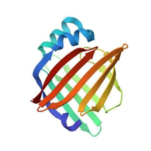
    3HK1

  • PubMed Abstract: 

    Molecular disruption of the lipid carrier AFABP/aP2 in mice results in improved insulin sensitivity and protection from atherosclerosis. Because small molecule inhibitors may be efficacious in defining the mechanism(s) of AFABP/aP2 action, a chemical library was screened and identified 1 (HTS01037) as a pharmacologic ligand capable of displacing the fluorophore 1-anilinonaphthalene 8-sulfonic acid from the lipid binding cavity. The X-ray crystal structure of 1 bound to AFABP/aP2 revealed that the ligand binds at a structurally similar position to a long-chain fatty acid. Similar to AFABP/aP2 knockout mice, 1 inhibits lipolysis in 3T3-L1 adipocytes and reduces LPS-stimulated inflammation in cultured macrophages. 1 acts as an antagonist of the protein-protein interaction between AFABP/aP2 and hormone sensitive lipase but does not activate PPARgamma in macrophage or CV-1 cells. These results identify 1 as an inhibitor of fatty acid binding and a competitive antagonist of protein-protein interactions mediated by AFABP/aP2.


  • Organizational Affiliation
    • Department of Biochemistry, Molecular Biology, and Biophysics, University of Minnesota, Minneapolis, Minnesota 55455, USA.

Macromolecule Content 

  • Total Structure Weight: 14.88 kDa 
  • Atom Count: 1,269 
  • Modeled Residue Count: 131 
  • Deposited Residue Count: 131 
  • Unique protein chains: 1

Macromolecules

Find similar proteins by:|  3D Structure
Entity ID: 1
MoleculeChains  Sequence LengthOrganismDetailsImage
Fatty acid-binding protein, adipocyte131Mus musculusMutation(s): 0 
Gene Names: adipocyteAp2Fabp4fatty acid binding protein 4
UniProt
Find proteins for P04117 (Mus musculus)
Explore P04117 
Go to UniProtKB:  P04117
Entity Groups
Sequence Clusters30% Identity50% Identity70% Identity90% Identity95% Identity100% Identity
UniProt GroupP04117
Sequence Annotations
Expand
Reference Sequence

Small Molecules

Ligands 1 Unique
IDChains Name / Formula / InChI Key2D Diagram3D Interactions
B64

Query on B64


Download:Ideal Coordinates CCD File
B [auth A]4-{[2-(methoxycarbonyl)-5-(2-thienyl)-3-thienyl]amino}-4-oxo-2-butenoic acid
C14 H11 N O5 S2
GJODSFZNKNHKML-PLNGDYQASA-N

Experimental Data & Validation

Experimental Data

  • Method: X-RAY DIFFRACTION
  • Resolution: 1.70 Å
  • R-Value Free:  0.195 (Depositor), 0.195 (DCC) 
  • R-Value Work:  0.175 (Depositor), 0.174 (DCC) 
  • R-Value Observed: 0.176 (Depositor) 
Space Group: C 2 2 21
Unit Cell:
Length ( Å )Angle ( ˚ )
a = 77.017α = 90
b = 94.113β = 90
c = 49.662γ = 90
Software Package:
Software NamePurpose
REFMACrefinement
PDB_EXTRACTdata extraction
CrystalCleardata collection
HKL-2000data reduction
HKL-2000data scaling
MOLREPphasing

Structure Validation

View Full Validation Report


Ligand Structure Quality Assessment 


Entry History 

Revision History  (Full details and data files)

  • Version 1.0: 2009-09-22
    Type: Initial release
  • Version 1.1: 2011-07-13
    Changes: Version format compliance
  • Version 1.2: 2023-09-06
    Changes: Data collection, Database references, Derived calculations, Refinement description